Chapter 17 Nonolfactory Rhinencephalon
Limbic System
The prosencephalon consists of the telencephalon and the diencephalon. The diencephalon is composed of the thalamus, epithalamus, metathalamus, subthalamus, and hypothalamus, with the third ventricle in the center. The telencephalon is the cerebrum, which consists of two cerebral hemispheres. The cell bodies of the neurons in the cerebrum are located in two general areas. One area is on the external surface of the gyri in the laminations of the cerebral cortex. The other area is deep to the surface in basal nuclei. The caudate nucleus, pallidum, and putamen are basal nuclei that function in the extrapyramidal system. The amygdaloid body and septal nuclei function mainly in the limbic system.
Another grouping of brain components is called the rhinencephalon, the “smell brain.” It consists of an olfactory component, the paleopallium (see Chapter 16), and a nonolfactory component, which is referred to as the limbic system. In the telencephalon, this includes the archipallium.
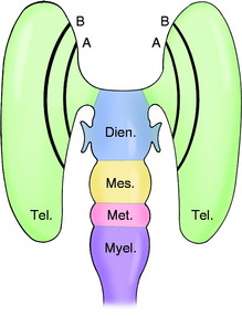
The term limbic system refers to the anatomic arrangement of the telencephalic neurons and tracts that are components of this system and are arranged primarily as two incomplete ring-like structures on the medial aspect of the cerebral hemisphere at its border with the diencephalon (Fig. 17-1). Limbus means edge or border, and these telencephalic structures form the border of the main mass of the cerebral hemisphere. The term has been expanded in usage to include the major nuclei and pathways in the rostral brainstem that are connected with these telencephalic structures anatomically and functionally (Box 17-1).
Box 17-1 Nonolfactory Rhinencephalon: Limbic System, Summary of Major Structures
ANATOMY
Telencephalon
The telencephalic components of the limbic system form two “cortical rings” at the border of the diencephalic-telencephalic junction (see Fig. 17-1). The inner ring consists of the amygdaloid body, the hippocampus, and its fornix. The outer ring consists of the cingulate gyrus and its cingulum and the septal area.
The amygdaloid body, one of the basal nuclei of the telencephalon, is a complex of nuclei located in the piriform lobe deep to the olfactory cortex (Fig. 17-2; see also Fig. 2-3). A projection pathway of the neurons in the amygdaloid nuclei, the stria terminalis, courses in the angle between the thalamus and the caudate nucleus. It forms an incomplete circle shaped like a C that progresses from the amygdaloid body in a caudal, then dorsal, and then rostral and ventral direction to terminate in the septal area and the rostral hypothalamus. A diagonal band courses on the ventral surface of the cerebrum, connecting the amygdaloid body with the septal area.
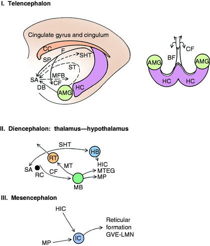
The hippocampus is a unique gyrus of the cerebrum that in lower mammals is found on the external surface of the cerebrum but in higher mammals has been rolled into the lateral ventricle and is not visible on the external surface. The hippocampus is shaped like a C. The hippocampus extends in a curve, starting from the amygdaloid body ventrally in each piriform lobe and progressing caudally and dorsally and then rostrally over the diencephalon. It forms part of the medial and dorsal wall of the lateral ventricle ventrally, and part of the medial and ventral wall of the lateral ventricle dorsally (see Fig 4 through 6). It lies adjacent to the lateral geniculate nucleus from which it is separated by meninges with cerebrospinal fluid. Dorsal to the caudal thalamus, the hippocampus of each cerebral hemisphere meets at the median plane, and a commissure is formed here, the hippocampal commissure. On the ventral surface of the brain, caudal to the piriform lobe, the hippocampus is covered superficially by the parahippocampal gyrus, which is bounded laterally by the caudal lateral rhinal sulcus and medially where it joins the hippocampus at the hippocampal sulcus (see Fig. 2-4). The parahippocampal gyrus is continued dorsally, dorsal to the corpus callosum, by the cingulate gyrus (see Fig 4 through 6).
Axons course to and from the hippocampus along its lateral side, forming the fimbria and the crus of the fornix. The two crura meet rostral to the hippocampal commissure and continue rostrally as the body of the fornix (see Figs. 17-2, 2-2, and 2-3). After coursing rostrally a short distance, the body of the fornix bends ventrally and dorsal to the rostral commissure. Two distinct cylindric columns emerge from the body on each side. Each column of the fornix splits at the rostral commissure. On each side, a small bundle of axons courses rostrally into the septal area, and a larger portion of the column courses caudal to the rostral commissure and ventrally through the hypothalamus beside the third ventricle to terminate in the mamillary body (see Figs. 2-3 and 2-4). The body of the fornix and the proximal portion of each column are attached dorsally to the corpus callosum by the septum pellucidum. The caudal portion of this septum is not visible unless the lateral ventricle is enlarged and the septum appears as a thin sheet between the body of the fornix and the corpus callosum. Rostrally, this septum contains neuronal cell bodies of the septal nucleus. The leptomeninges extend rostrally between the diencephalon and the hippocampi and body of the fornix to the level of the interventricular foramen. The columns of the fornix that course caudal to the rostral commissure form the rostral border of this foramen on each side. The caudal border of this foramen is the portion of choroid plexus that is continuous from the third ventricle to the lateral ventricle. Here, the rostral extent of the leptomeninges between the diencephalon and telencephalon is associated with this choroid plexus.
The cingulate gyrus consists of cerebral cortex and its corona radiata, the cingulum. It is located dorsal to the corpus callosum and is continuous caudally with the parahippocampal gyrus and rostrally with the septal area (see Fig 2-2 through 2-6). The cingulum is a long association tract consisting of longitudinal axons in the white matter (corona radiata) of the cingulate gyrus (see Fig. 17-2). These axons course from the parahippocampal gyrus, located caudally, to the septal area and frontal lobe gyri located rostrally.
The septal area consists of the subcallosal area, which is the cerebral cortex ventral to the genu of the corpus callosum and the septal nuclei. The septal nuclei are a collection of neuronal cell bodies in the rostral septum pellucidum that bulges into the medial side of the lateral ventricle. This is dorsal and just rostral to the bend in the body of the fornix, where it forms the columns of the fornix. The septal area connects with the hippocampus by way of the adjacent columns of the fornix; with the amygdaloid body through the diagonal band and stria terminalis; and with the habenular nucleus via the stria habenularis thalamus (see Fig. 2-3). The medial forebrain bundle courses caudally from the septal area into the hypothalamus. By way of this pathway, limbic system efferents can influence the hypothalamic centers that control the activity of the general visceral efferent (GVE) system. The function of the limbic system involves visceral motor activation. Hypothalamic nuclei serve in the upper motor neuron system that regulates the GVE system. These hypothalamic nuclei receive numerous limbic system efferents.
Stay updated, free articles. Join our Telegram channel

Full access? Get Clinical Tree


