18 No clinical signs of root resorption
ORAL EXAMINATION – CONSCIOUS
The cat allowed conscious examination of the head and mouth, which identified the following:
ORAL EXAMINATION – UNDER GENERAL ANAESTHETIC
In summary, examination under general anaesthesia identified the following:
RADIOGRAPHIC FINDINGS
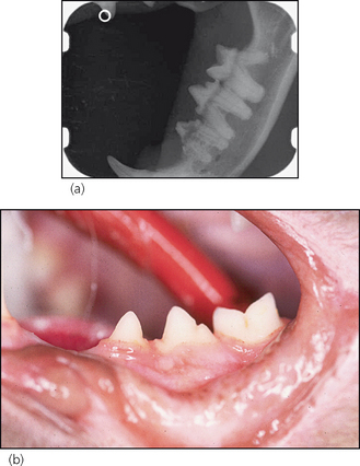
Figure 18.1 Radiograph (a) and lateral photograph (b) of the left lower premolars and molar.
THEORY REFRESHER
Visual inspection and tactile examination with a dental explorer will only identify end-stage lesions, i.e. when the process involves the crown and has resulted in an obvious cavity. Radiography will identify lesions that are localized to the root surfaces within the alveolar bone (as in this case), which would not be detected by clinical methods. Moreover, it is only with the aid of radiography that the extent of a resorptive process can be identified. Selection of the best treatment option thus depends on radiography. In fact, a series of full-mouth radiographs (the technique is covered in Chapter 4) is recommended for all cats presented for dental therapy. If taking a series of full-mouth radiographs is not possible, e.g. financial restrictions, then take one view of each mandibular premolar/molar region. The mandibular third premolars are the most commonly affected teeth and it has been shown that, in nine out of 10 cats with resorptive lesions, the process will be identified on these two views. If radiographs show resorption of these teeth, then a full-mouth series must be taken.
It remains a matter of debate as to whether odontoclastic resorptive lesions (ORLs) cause discomfort or pain to the affected individual. Based on the fact that pulpal inflammation occurs late in the disease process, it seems likely that lesions that are limited to the root surfaces and do not communicate with the oral environment are asymptomatic. However, once dentine destruction has progressed to such an extent that the process invades the pulp and/or a communication with the oral cavity has been established (when the enamel has been resorbed or it has fractured off to reveal the dentine to the oral cavity), then discomfort and/or pain are likely. Some cats may show clinical signs indicating oral discomfort or pain, e.g. changes in food preferences (soft rather than hard diet) and reduced food intake, but many cats do not.


