, Patricia A. Noguera1 and Trygve T. Poppe2
(1)
Marine Scotland Science, Aberdeen, UK
(2)
Norwegian School of Veterinary Science, Oslo, Norway
Abstract
A neoplasm represents an altered process involving abnormal and uncontrolled growth of cells that is usually detrimental to the host. Neoplasm are classified as benign or malignant and named according to the features of differentiation recognised through histological examination, thus reflecting the tissue of origin. Neoplasia can arise following exposure to certain chemicals, heavy metals, ionizing radiation, chronic inflammation, ultraviolet radiation, certain viruses and pollution. The chapter covers a selection of reported neoplastic conditions affecting fish although overall, many of the cause(s) have not been determined.
Keywords
NeoplasiaSalmonTroutA neoplasm is an altered process involving abnormal and uncontrolled growth of cells that is usually detrimental to the animal. They are named according to the features of differentiation recognised through histological examination, thus reflecting the tissue of origin. In this context, neoplasm and tumour are interchangeable terms in current medical usage. The abnormal cell type may continue to increase even after the initiating mutagen is no longer present and generally more than one mutation is necessary for oncogenesis. Neoplasia can arise following exposure to certain chemicals, heavy metals, ionizing radiation, chronic inflammation, ultraviolet radiation, certain viruses and pollution. They are classified as benign or malignant (i.e. to include anaplasia, frequent mitotic figures and infiltration of neutrophils), but overall many of the cause(s) have not been determined. Examples of neoplasia representing a range of tissue origins are discussed.
12.1 Papilloma
Atlantic salmon papillomatosis is an epidermal benign neoplasia on the skin and scales of salmon parr in their second summer, but also occasionally of young adult fish (smolts and grilse) which have adapted to sea water.
The growths appear as single or multiple with a smooth to nodular texture, and range from white to brown or pink. Each papilloma may vary from a few millimetres to complex growths of up to 40 mm (Fig. 12.1). In heavily affected fish more than half of the body surface may be covered. Histologically, it is a plaque-like proliferation of the epidermis (Fig. 12.2). Each plaque is a stratified squamous epithelium with supporting stroma, hyperplastic epidermal cells containing prominent nucleoli with numerous atypical mitotic figures. There is usually little dermal involvement. The number of mucous cells is reduced and the basement membrane absent or indistinct. The papilloma is relatively harmless to the fish and eventually detaches allowing the skin to heal.
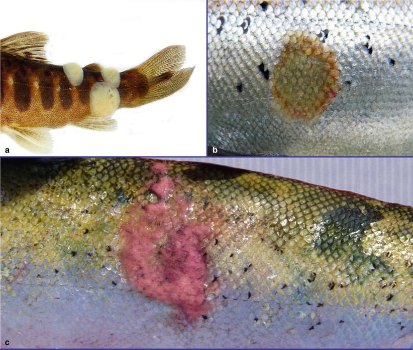
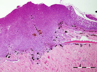
Fig. 12.1
(a) Papillomatosis in wild Atlantic salmon parr, alcohol-preserved specimen. (b) Healing papillomatosis in wild adult Atlantic salmon. (c) Papillomatosis in farmed adult Atlantic salmon. Note haemorrhage around margins
Fig. 12.2
Section showing papillomatosis in Atlantic salmon parr. Normal skin with mucous cells to the right. Bar = 500 μm
Electron microscopy studies of papillomas have indicated that virions with herpesvirus morphology are sometimes associated with the growth. Attempts to isolate this agent in culture have failed and therefore, the cause (or possibly causes) of papillomatosis remains unknown.
12.2 Chondroma
Single, firm, ovoid, white, smooth growths involving the branchial cartilage have been reported in salmonids and diagnosed as a benign cartilaginous neoplastic growth or chondroma (Fig. 12.3). These are encapsulated with a lobular growing pattern that originates from the filament cartilage, possibly as an ingrowth of the surface covering. Normal squamous, or sometimes hyperplastic epithelium covers the surface of the mass with some occasional invaginations into the underlying stroma with increased mucous cell activity. Within the dermis, there is hypertrophy and some hyperplasia. Many of these growths contain a number of cystic spaces beneath the epidermis and are surrounded by a loose fibrous matrix of adipose tissue containing strands of immature cartilage (Fig. 12.4). Melanin granules are evenly spread throughout the dermis with no evidence of inflammation, infiltrative growth or distant metastasis. Chondrocytes are infrequent with no mitotic figures.
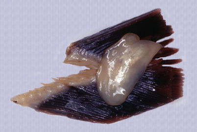
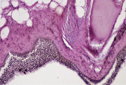
Fig. 12.3
Chondroma in the gill of rainbow trout
Fig. 12.4
Transverse section showing gill chondroma in rainbow trout. Lillie’s allochrome stain. Low magnification
The absence of infectious agents coupled with the rarity of chondromas in populations indicates that these growths are natural occurrences, hence the cause or causes are considered to be spontaneous in nature. The topographic localization and histological features are used for diagnosis.
12.3 Pigment Cells
12.3.1 Melanoma
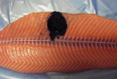
Pigment cell neoplasia is occasionally reported in salmonids, with melanomas being the most common type from this group. Both wild and farmed stock may be affected. The mature growths are generally raised, soft, black-pigmented areas which are visible on the body surface and underlying muscle (Fig. 12.5). The neoplasia shows invasion by melanomacrophages in varying degrees of differentiation, with fibrous deposition (Fig. 12.6). Metastasis has been recorded. Gross observations and the cellular detail described from stained histological sections are used to confirm the diagnosis.