Navicular Disease

Chapter 30 Navicular Disease


Pathophysiology of Navicular Disease


Navicular disease is a chronic forelimb lameness associated with pain arising from the distal sesamoid or navicular bone. It is well recognized that in association with advanced navicular disease, fibrillation of the dorsal aspect of the deep digital flexor tendon (DDFT), with or without adhesion formation between the tendon and the navicular bone, is a common feature. Recent clinical studies using magnetic resonance imaging (MRI)1 and post mortem studies2,3 have demonstrated that there may also be abnormalities of closely related structures, including the collateral sesamoidean ligaments (CSLs), the distal sesamoidean impar ligament (DSIL), and the navicular bursa. These structures and the navicular bone are called the podotrochlear apparatus. For the purposes of this chapter, this complex of degenerative changes will be referred to as navicular disease. Primary injury of the DDFT is considered to be a separate condition,4 which may have a different etiopathogenesis (see Chapter 32).


Although historically considered to be a single disease, given the variety of clinical presentations it is likely that there are a number of different clinical conditions, of different etiologies, that give rise to pain in the podotrochlear apparatus. It is difficult to conceive a single disease that can result in an insidious onset, slowly progressive bilateral forelimb lameness, or an acute onset, relatively severe unilateral forelimb lameness, each with a variety of different radiological manifestations and with some horses never developing radiological changes. It is curious that sometimes clinical signs become apparent in young horses just commencing work, whereas more typically lameness is seen in mature riding horses. It is also seen in horses with vastly different distal limb conformation. It is a common condition in Quarter Horses, which tend to have narrow, upright, boxy feet, small relative to their body size, as well as in European Warmblood horses, many of which have relatively tall narrow feet. It is also common in Thoroughbred horses, which frequently have rather flat feet, with low collapsed heels, often associated with dorsopalmar foot imbalance. Recent evidence suggests there is a heritable tendency toward the development of navicular disease in Dutch and Hanoverian Warmblood horses.5-7


The factors causing pain in the navicular apparatus, and therefore lameness, are poorly understood. Experience with both radiography and MRI suggests that lesions in both the navicular bone and closely related structures are likely to predate the onset of lameness in some horses.8 In some horses, a trigger factor apparently promoting pain and lameness has been a period of enforced rest for an unrelated cause.


There are no good epidemiological studies investigating risk factors for the development of navicular disease. Therefore information is largely anecdotal. The frequency of occurrence of navicular disease appears to vary between breeds. Quarter Horses,9 Warmblood horses,10 and Thoroughbred cross horses11 have a relatively high incidence, whereas the occurrence and/or recognition in some breeds, such as the Finnhorse, Arab,12 and Friesian, is relatively low.


Biomechanical Considerations


From a biomechanical perspective the podotrochlear apparatus comprising the navicular bone, the CSLs, the DSIL, and the navicular bursa, together with the DDFT, and the distal digital annular ligament are integrally related. The navicular bone, which articulates with the middle and distal phalanges, provides a constant angle of insertion and maintains the mechanical advantage of the DDFT, which exerts major compressive forces on the distal third of the bone. Contact studies between the phalanges in isolated limbs have demonstrated that the greatest forces are applied in the propulsion phase of the stride. This occurs during extension of the distal interphalangeal (DIP) joint, with increased pressure of the DDFT on the palmar aspect of the navicular bone, increased contact between the navicular bone and the middle phalanx, and increased tension in the CSLs.13,14 Tension in the DDFT and the distal digital annular ligament promotes stability of the DIP joint. Forces may be altered by foot conformation: in a horse with “weak” heels there is greater extension of the DIP joint compared with a horse with “strong” heels, which results in increased pressure concentrated on the distal aspect of the navicular bone.15


Compressive forces and stress on the navicular bone were compared in clinically sound horses and horses with navicular disease.16 Although the mean peak force and stress were similar, the force and stress in the horses with navicular disease were approximately double early in the stance phase of the stride. This early peak stress resulted in a much higher loading rate of the navicular bone in the navicular disease group. The difference in loading patterns was associated with an increased force in the DDFT in the early and mid-stance phases, probably because of increased contraction of the deep digital flexor (DDF) muscle. Contraction of the DDF muscle may result in toe-first ground contact, seen in some horses with navicular disease. It is suggested that pain associated with the navicular bone may result in a positive feedback by increasing the force in the DDFT to avoid heel-first landing, and hence paradoxically increasing the compressive force on the navicular bone. This hypothesis is supported by reduction in peak forces on the navicular bone throughout the stance phase in horses with navicular disease after perineural analgesia of the palmar digital nerves.17


Although low, collapsed heel conformation has anecdotally been associated with navicular disease, a recent study18 in Irish draught cross-type horses showed no correlation between the peak force exerted on the navicular bone by the DDFT and the conformation of the hoof capsule, contrary to the earlier results.15 However, a 1-degree decrease in angle of the solar border of the distal phalanx resulted in a fourfold increase in peak force on the navicular bone.18 There was no correlation between the angle of the solar border of the distal phalanx and the degree of heel collapse.


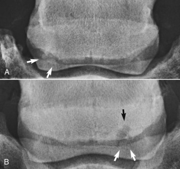
The shape of the navicular bone may be determined at birth, and this may influence the biomechanical forces subsequently applied to the bone (Figure 30-1) and hence influence the risk of development of navicular disease.6,19 Finnhorses and Friesian horses tend to have a straight or convex contour of the proximal articular border of the navicular bone and rarely develop navicular disease. There is a much higher incidence of navicular disease in the Dutch Warmblood breed, and horses in which the proximal articular margin is concave or undulating appear to be at highest risk of development of the disease.5,6


Histopathological Studies


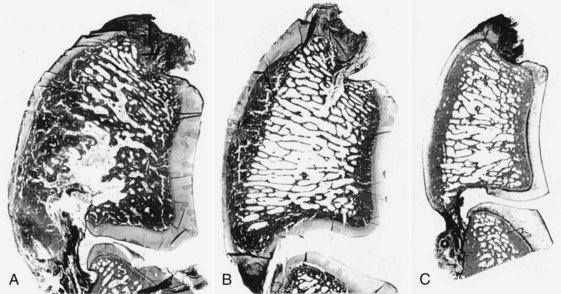
Navicular disease has not been reproduced experimentally; therefore all proposed etiologies remain speculative. Earlier theories suggesting a vascular etiology with arteriosclerosis20 or thrombosis, resulting in ischemia within the navicular bone,21 have largely been rejected because of failure to identify ischemic bone or thrombosis, failure to reproduce clinical signs or pathological changes by occluding blood supply to the bone, and expanding evidence demonstrating increased bone modeling.22-25 Post mortem studies to date have focused principally on horses with long-term, chronic disease, generally with advanced radiological abnormalities, reflecting the end stage of a disease complex. These studies identified striking similarities between the pathological features of navicular disease and osteoarthritis (Figure 30-2) in both people and horses.24,25


Studies of aging changes in the navicular bone of normal immature and mature horses suggested that there is a degenerative aging process similar to that seen in joints.24 However, a more recent study investigating not only the navicular bone but also the DDFT, CSLs, DSIL, and navicular bursa demonstrated no age-related differences between mature horses with no history of foot-related lameness ranging from 4 to 15 years of age.2,3 This suggests that there may be an individual susceptibility to degenerative change. Nonphysiological biomechanical factors may promote this susceptibility to degenerative change.16,24,26


The explanation for pain and lameness in horses with no detectable radiological change has been poorly investigated by post mortem studies. However, recent clinical experience with MRI has indicated that many horses with evidence of increased modeling of the navicular bone based on increased radiopharmaceutical uptake (IRU) detected using nuclear scintigraphy do have pathological abnormalities of the navicular bone detectable using MRI, with or without concurrent changes in the DDFT, CSLs, DSIL, and navicular bursa.27,28


Degenerative changes in the fibrocartilage on the palmar aspect of the navicular bone occur principally in the distal half of the bone, especially centered around the sagittal ridge in both sound and lame horses.2 In horses with navicular disease there is a greater degree of fibrocartilage damage, which may extend into the subchondral bone. Partial-thickness loss of fibrocartilage in this location was one of the most common lesions significantly associated with navicular disease in one study.25 It is likely to represent some of the earliest pathology of one form of this disease but remains difficult to identify in vivo, even with the use of MRI.8 Degenerative change of the spongiosa is generally only seen dorsal to extensive fibrocartilage damage. Physiological forces result in adaptive remodeling of the subchondral bone in immature horses, with cortical thickening.24 Nonphysiological forces may result in focal fibrocartilage and/or flexor cortex damage, with adjacent subchondral sclerosis dorsal to it, associated with thickening of trabeculae and focal areas of lysis. There may also be edema, congestion, and fibrosis of the marrow stroma within the medullary bone, which may result in a cystlike lesion.


Concurrently there may be fibrillation of the dorsal surface of the DDFT, which may predispose to adhesion formation between the DDFT and regions of partially or fully eroded fibrocartilage on the palmar aspect of the navicular bone. Whether lesions in the DDFT are primary or secondary to preexisting damage of the fibrocartilage currently remains open to debate. However, recent post mortem evidence suggests that there may be non–age-related degenerative vascular and matrix changes in the dorsal aspect of the DDFT in both lame and clinically normal horses.3 Although it has been suggested that vascular occlusion and matrix changes in the DDFT may be age-related,25 the results of a recent study showed that the severity of these changes was greater in horses with palmar foot pain than in age-matched control horses.3 Minor fibrillation of the dorsal aspect of the DDFT was seen in both lame and control horses, whereas deep sagittal splits were only seen in lame horses. Complete occlusion of blood vessels, replacement of normal tendon architecture by focal fibroplasia, and areas of fibrocartilaginous metaplasia were common in the lame horses. As these changes are predominantly seen in the intratendonous septa, there is a strong possibility that they predispose to the development of sagittal splits in the dorsal surface of the tendon along these septal planes. Sharp edges of splits in the DDFT extending from the dorsal surface may cause ulceration of the fibrocartilage of the navicular bone and thus predispose to lesions extending into the medulla.


There is an association between changes of the flexor aspect and distal and proximal borders of the navicular bone.2 Similar types of change occur at the proximal and distal aspects but tend to be more extensive distally. Enlarged synovial invaginations are the result of recruitment and activation of osteoclasts following the course of the nutrient vessels into the spongiosa. This may be associated with local medullary osteonecrosis and the presence of foci of fibrocartilaginous metaplasia and/or entheseous new bone close to the interface between the DSIL and the navicular bone.


Aging changes were described in the articular cartilage of the navicular bone and the opposing face of the distal phalanx.29 There was loss of proteoglycan and tidemark advancement, which was thought to reflect excessive shear stress in the zone between the calcified and noncalcified articular cartilage. A greater number of tidemarks were seen in horses with clinical signs of navicular disease than normal horses of similar age. However, a more recent study failed to identify significant age-related changes, and low-grade degenerative changes in the articular cartilage were common in both control horses and those with navicular disease.2


Observations from Nuclear Scintigraphy and MRI


Previous studies using tetracycline labeling of bone22 and scintigraphic studies27,30 have indicated that there is evidence of increased bone turnover in association with some forms of navicular disease, even in the absence of radiological abnormalities of the bone. IRU predominantly reflects increased osteoblastic activity31 but is not synonymous with either pain or lameness.32 IRU may reflect a functional adaptation to foot conformation and the biomechanical forces on the navicular bone. Comparison between scintigraphy and MRI has demonstrated that many horses with focal moderate or intense IRU have abnormalities of the navicular bone detectable using MRI.27 However, scintigraphy can also produce false-negative results, indicating that pathological abnormalities of the navicular bone are not always associated with increased osteoblastic activity.


A comparison of MRI findings in control horses with no history of foot-related pain and horses with chronic palmar foot pain showed significant alterations of the podotrochlear apparatus in the lame horses.33 A comparative MRI and post mortem study showed good correlation between the lesions identified using MRI and histopathological findings.34 Clinical experience with MRI in horses with foot pain provides support for the progression of lesions as outlined above and has demonstrated some earlier lesions than those investigated post mortem.8


However, a group of horses has also been identified with no detectable abnormalities of the flexor fibrocartilage or cortex but with diffuse abnormalities of the medulla characterized by increased signal intensity in fat-suppressed magnetic resonance (MR) images. Post mortem examination of several such horses revealed evidence of early fat necrosis with a moth-eaten appearance of the trabeculae, with necrosis of bone edges. This may have a different etiopathogenesis.


Hyperintense signal in the medulla of the navicular bone has been ascribed to the presence of edema in the marrow spaces,35 but this was not validated post mortem. Further research is required to determine the true causes of this phenomenon. In our studies, mild or moderate focal or generalized increased signal intensity in fat-suppressed MR images was associated with trabecular thinning and widened intertrabecular spaces.34 High-intensity increased signal associated with irregular decreased signal intensity in T1- and T2-weighted images was associated with generalized osteonecrosis and fibrosis, with irregular trabeculae, adjacent adipose tissue edema, and prominent capillary infiltration. A recent post mortem study of feet with advanced radiological abnormalities of the navicular bone demonstrated that increased signal intensity in fat-suppressed images correlated with areas of degenerate adipose tissues, hemorrhage or replacement by fibrocollagenous material, or fluid-filled cystic spaces.36


In some other horses, fluid-filled osseous cystlike lesions were seen in the distal aspect of the bone, apparently separate from synovial invaginations, and not associated with any detectable abnormality of the flexor aspect of the bone. Such lesions have not yet been characterized histologically, and their etiology remains speculative, although they may be associated with lesions of the DSIL.


Occasionally horses have been identified with new bone on the palmar aspect of the navicular bone, centered on the sagittal ridge. The cause of this is currently unknown.


Entheseous Changes


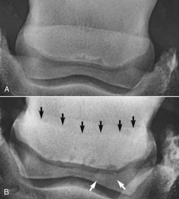
The presence of entheseous new bone on the proximal border of the navicular bone, reflecting previous insertional desmopathy of the CSL, is well documented radiologically37,38 and at post mortem examination25,37 in both clinically normal horses and horses with navicular disease. Its clinical significance remains uncertain, although more extensive new bone in this location tends to be associated with other signs of navicular disease.37,38 Recent experience with MRI has confirmed these findings.1 Rarely, an avulsion fracture is identified at the insertion of the CSL into the navicular bone.8,39 Mineralized and osseous fragments (Figure 30-3) in the DSIL have also been recognized in both normal horses and in horses with navicular disease, and their clinical significance remains difficult to determine. Fragments were unusual in sound horses undergoing prepurchase radiographic examination,40 although their true incidence may be underestimated by radiographic examination compared with MRI or computed tomography (CT). In two post mortem studies, fragments associated with a defect in the distal margin of the navicular bone were more common in horses with navicular disease than in age-matched controls.2,25 This has also been my clinical experience.


Fibrocartilaginous metaplasia in the body of the DSIL was more extensive in horses with navicular disease compared with age-matched control horses.2 However, no significant differences between groups were seen in the CSLs.


Aging changes have been seen in the region of insertion of the DSIL and DDFT, with a change in fibroblast shape and an increase in proteoglycans.14 The functional significance of this is not yet known. Evidence of inflammation was recently recognized histologically at the intersection of the DSIL and DDFT in horses with clinical signs of navicular syndrome.29,41 Changes reflecting “abnormal stress” at the insertion of the DSIL and DDFT have been demonstrated in horses with poor foot conformation.29 This region is rich in sensory nerve endings, with many arteriovenous complexes that are damaged in horses with navicular disease.42


Clinical experience with MRI has demonstrated that structural abnormalities of the DSIL are often seen in association with abnormal modeling of the distal palmar aspect of the navicular bone, with focal increased signal in fat-suppressed MR images.1,28 It has been suggested that this focally increased signal in the distal aspect of the navicular bone at the origin of the DSIL on fat-suppressed images may represent an important early event in the etiopathogenesis of navicular disease.33 Increased signal intensity may also be seen in the navicular bone close to the insertion of the CSLs in fat-suppressed images.1,28 In some horses there is a linear band of increased signal intensity in fat-suppressed images, extending through the middle third of the navicular bone, from the insertion of the CSL to the origin of the DSIL. Abnormalities of the CSL have also been associated with concurrent abnormalities of the navicular bone. Based on clinical experience, it seems that these lesions may be the result of abnormal stresses at the attachments of the CSL and DSIL on the navicular bone, and this may reflect a different mechanism of navicular disease development.8


Although endosteal irregularity at the insertion of the DSIL on the distal phalanx may be seen in both horses with and without foot pain,33,34 in some lame horses there is evidence of insertional desmopathy, characterized by enthesophyte formation, axial cortical disruption, osseous cystlike leions, and/or increased signal intensity in the bone at this site in fat-suppressed images, reflecting bone edema or necrosis.8


Diagnostic Considerations


Clinical Signs


Pain when the horse is standing at rest may be a feature of navicular disease; however, this is a variable finding, and some clinically normal horses habitually point one or both front feet. A horse that has resting pain associated with navicular disease may stand pointing one front foot, sometimes alternating between feet. This is also a feature of horses with primary lesions of the DDFT. Alternatively, an affected horse may pack bedding under the heel or have a tendency to sit on a manger to relieve pressure on the palmar aspect of the foot. Navicular disease is recognized in horses with a wide variety of foot shapes: narrow, boxy upright feet of the Quarter Horse and low, collapsed heels typical of many Thoroughbreds. Navicular disease often is seen in association with poor mediolateral or dorsopalmar foot balance. If lameness is consistently worse on one forelimb, the feet may become asymmetrical in shape, with the lamer foot narrower with a taller heel.


The digital vessels sometimes are palpably enlarged, but this is an inconsistent, nonspecific finding. The response to hoof testers applied to the frog region is often negative,22,49,50 although other authors51 have described a positive response as a fairly consistent feature of navicular disease. Pain may be elicited at the toe if the horse has been repeatedly overloading the toe, resulting in subsolar bruising. Differences in clinical signs observed by different clinicians may reflect genuine differences in horse populations in different geographical locations. Distention of the DIP joint capsule is sometimes seen in association with navicular disease but not invariably so. Increased intraarticular pressure (>40 mm Hg) is said to be associated with navicular disease,52 although in my experience there is considerable variability in the degree of DIP joint distention and pressure in both normal and lame horses.


Lameness is sometimes apparent when the horse is moving on a hard surface in straight lines. It may fluctuate in degree within an examination period or between examinations performed on different days. The horse may show a tendency to stumble associated with an altered foot placement. Overt unilateral lameness may be evident, but in some horses there is only marginal shortening of stride and reduced lift to the stride, which may be difficult to detect if the horse was not previously known to the observer. The horse may move better on a soft surface, even in circles. No clinical signs are detectable in some horses when examined moving in hand on a hard surface. Lameness generally is accentuated if the horse moves in circles on a hard surface, especially with the lame limb on the inside of the circle. In some horses, lameness is apparent only under these circumstances. Less commonly lameness is accentuated when the lame limb is on the outside of a circle. Sometimes lameness cannot be detected unless the horse is ridden, when it may move in a slightly stiff, “flat,” and restricted fashion. Recognition of this requires previous knowledge of the horse, knowledge of the expected quality of movement of a horse of that type, or respecting the opinion of the rider that the horse has “lost some action.” A vast difference may be detected by desensitizing both front feet. This may be easier to appreciate when riding the horse than watching it.


The response to distal limb flexion is extremely variable. Many horses with navicular disease show a transient, mild increase in lameness.22,49,50 Resistance to flexion or marked accentuation of lameness after flexion is unlikely to reflect navicular bone pain. Distal limb flexion of one forelimb may result in increased lameness in the contralateral forelimb because of increased loading of the podotrochlear apparatus, but the same response can also be seen in horses with primary injuries of the DDFT. Elevation of the toe of the foot on a wedge or wooden board, with the contralateral limb picked up, resulting in extension of the DIP joint, may increase lameness, but this response is neither consistent nor pathognomonic for navicular disease.


If the horse’s foot conformation is poor and the feet are not trimmed and shod optimally, it is worthwhile improving the trimming and shoeing and reassessing the lameness after several weeks. If the lameness has markedly improved, it is unlikely to reflect navicular disease.


Jun 4, 2016 | Posted by in EQUINE MEDICINE | Comments Off on Navicular Disease

Full access? Get Clinical Tree

Get Clinical Tree app for offline access