Mycotoxins and mycotoxicoses

Chapter 44


Mycotoxins and mycotoxicoses


Although moulds can grow on a wide range of organic matter including growing crops and stored feed, those of greatest significance in veterinary medicine produce secondary metabolites which are toxic for many animal species and man in low concentrations. Fungal toxins are referred to as mycotoxins and the diseases they produce are termed mycotoxicoses. Production of mycotoxins occurs as a result of normal fungal metabolism. No specific role for these metabolites in the life cycle of the fungus has been demonstrated. Some mycotoxicoses such as ergotism have been known since the Middle Ages while many other diseases arising from the ingestion of mycotoxin-contaminated pasture or feed have been recognized only in recent decades.


Mycotoxicoses are not infections but are acute or chronic intoxications produced by toxic metabolites of fungal origin. Many of the toxigenic fungi are widespread throughout the world and over 100 known species are capable of elaborating mycotoxins. Many of these fungi belong to the genera Aspergillus, Fusarium and Penicillium. More than one fungal species may produce the same mycotoxin while individual moulds may produce two or more different mycotoxins. Because taxonomic classification of fungi is based almost exclusively on morphological rather than physiological considerations, there is frequently little correlation between the pattern of toxin production by particular fungi and their phylogenetic classification. Toxin production only occurs under specific conditions of moisture, temperature, suitability of substrate and appropriate oxygen tension. The optimum conditions for toxin production are relatively specific for each fungus. Fusarium sporotrichoides elaborates its toxin at freezing temperatures while Aspergillus flavus requires a temperature of 25°C. Only some strains of a single species such as Aspergillus flavus have the ability to produce toxins, even under favourable conditions. Some strains of fungi have a distinct preference for certain substrates and consequently there may be a regional prevalence of particular mycotoxicoses depending on the types of crops or pasture cultivated in the region.


The susceptibility of different crops to mould infection is governed by the presence of suitable substrates. The seed or kernel may be the preferred target of some fungi because of the ready availability of carbohydrates, while the fibrous part of the plant with its high cellulose content may be invaded by other fungi capable of using this substrate. Damage to the seed coat by insects, mechanical harvesting, severe frost or other factors may predispose crops to fungal attack. Insects may also serve as carriers of fungal spores.


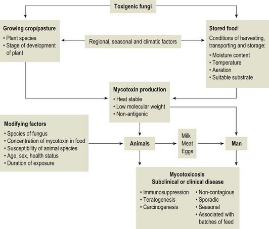
Mycotoxicoses are diseases in which many factors interact. Animals vary widely in their susceptibility to mycotoxins. Younger animals tend to be more susceptible than adults. There is also considerable variation in species and individual susceptibility. The conditions which favour mycotoxin production and the factors which influence the severity of mycotoxicoses are summarized in Figure 44.1. Since mycotoxins are most likely to be concentrated in highest amounts in stored feeds, groups of animals at risk include poultry, pigs, dairy and feedlot cattle fed on contaminated feed. In some countries, particularly New Zealand, mycotoxicoses such as ‘facial eczema’ are associated with standing pasture and accordingly prevailing climatic conditions determine the occurrence of disease.


Characteristics of Mycotoxins


As mycotoxins are low-molecular-weight, non-antigenic substances in their naturally occurring forms, acquired immunity does not occur following exposure. Many of the major mycotoxins are heat-stable and consequently can retain their toxicity after processing temperatures used for pelleting or other milling procedures. Each fungal toxin if present in the diet at a sufficient concentration usually affects specific target organs or tissues. Secondary mycotoxic disease may be more difficult to recognize because low levels of toxin intake may not result in a specific mycotoxicosis but in a heightened susceptibility to intercurrent infections due to immunosuppression. Some of the more important characteristics of mycotoxins are presented in Box 44.1.


Mycotoxicoses


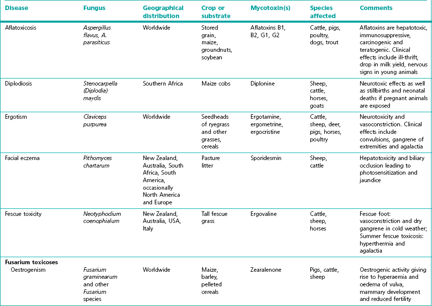
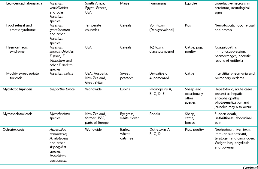
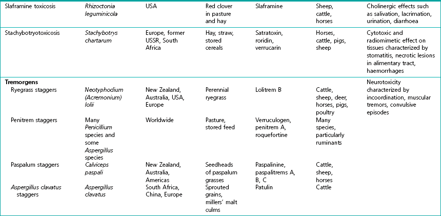
The severity of mycotoxicoses in animals and their clinical recognition is determined by many factors including the species of toxigenic fungus, the concentration of mycotoxin in the food, the age, sex and health status of the exposed animal, the target organ or tissue affected and the duration of exposure to contaminated feed. The features which characterize mycotoxic diseases include sporadic and seasonal occurrence, lack of transmissibility, association with certain batches of stored food or particular types of pasture, and disappointing response to drug treatment effective against infectious diseases (Box 44.2). Clinical and laboratory procedures may demonstrate pathological changes in the animal, characteristic of a particular intoxication. Clinical diagnosis can be complicated by the presence of a number of toxigenic fungal species on a food source. Diagnosis of a mycotoxicosis requires the demonstration of biologically effective concentrations of the fungal toxin in the feed available to the animal, or in the animal’s tissues, secretions or excretions. Some advances have been made in the application of molecular techniques for the detection of toxigenic fungi (Seifert & Levesque 2004, Mule et al. 2005). Table 44.1 summarizes the principal features of those mycotoxicoses which can be recognized clinically. The level of subclinical and chronic disease associated with the consumption of fungal toxins in feed is difficult to quantify but is certainly of economic and public health significance.


Aflatoxicosis


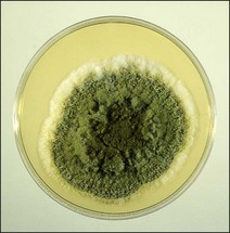
Aflatoxins are a group of approximately 20 related toxic compounds produced by some strains of Aspergillus flavus (Fig. 44.2), Aspergillus parasiticus and a number of other Aspergillus species during growth on natural substrates including growing crops and stored food. These fungi are ubiquitous, saprophytic moulds which grow on a variety of cereal grains and foodstuffs such as maize, cottonseed and groundnuts. About half of the strains of A. flavus and A. parasiticus are toxigenic under optimal environmental conditions. High humidity and high temperatures during preharvesting, harvesting, transportation and storage, as well as damage to field crops by insects, drought and mechanical injury during harvesting favour the growth of A. flavus and toxin production. Although other fungi such as Penicillium species and Rhizopus species, are capable of producing aflatoxins, their relevance to livestock production has not yet been established. The name ‘aflatoxin’ derives from Aspergillus(a-), flavus (fla-) and toxin.


One of the first, well-documented outbreaks of aflatoxicosis occurred in East Anglia, England in l960 when more than 100,000 turkey poults died of an unknown disease (‘turkey X disease’). Subsequently, it was demonstrated that these birds died from a toxin present in pelleted feed which formed a major part of their diet. A shipment of groundnut meal containing aflatoxin had been used as a protein supplement in the turkey rations. Examination of the incriminated groundnut meal revealed the presence of mould mycelia and thin-layer chromatography showed the presence of several compounds which fluoresced under ultraviolet light. The fungus was identified as Aspergillus flavus and the toxic metabolites were called aflatoxins. Since that time, numerous outbreaks of aflatoxicosis have been described worldwide.


Aflatoxins


Aflatoxins are a group of related difuranocoumarin compounds with toxic, carcinogenic, teratogenic and mutagenic activity. The four major aflatoxins are B1, B2, G1 and G2. Aflatoxin B1 (AFB1) is the most commonly occurring and also the most toxic and carcinogenic member of the group. These mycotoxins are named according to their position and fluorescent colour on thin-layer chromatograms, when viewed under ultraviolet light. AFB1 and AFB2 produce a blue and AFG1 and AFG2 a green fluorescence. Most of the other aflatoxins are metabolites formed endogenously in animals after ingestion or administration of aflatoxins. Aflatoxins are stable compounds in food and feed products and are relatively resistant to heat. They retain much of their activity after exposure to dry heat at 250°C and moist heat at 120°C but may be degraded by sunlight. They have a low molecular weight and are nonantigenic in their native state.


When growing in maize, A. flavus usually produces B1 and B2 aflatoxins, while A. parsiticus produces all four of the major aflatoxins. On soybeans, only low concentrations of AFB1 are produced by both species. Mould growth and toxin formation require a moisture content of the substrate greater than 15%, a temperature of at least 25°C and adequate aeration. Toxin formation can occur in a matter of hours when favourable conditions exist.


Biological effects of aflatoxins


The toxic effects of aflatoxins are dose-, time- and species-dependent. Mature ruminants are less susceptible to the effects of mycotoxins than young animals and monogastric animals. The toxins are absorbed from the stomach and metabolized in the liver to a range of toxic and nontoxic metabolites which are then excreted in urine and milk. The major biological effects of aflatoxins include inhibition of RNA and protein synthesis, impairment of hepatic function, carcinogenesis and immunosuppression. AFB1 is bioactivated in the liver to a highly reactive intermediate compound which reacts with various nucleophiles in the cell and binds covalently with DNA, RNA and protein. After deliberate administration of AFB1 there is marked interference with protein synthesis at the translational level which seems to correlate with disaggregation of polyribosomes in the endoplasmic reticulum. Many of the toxic responses observed in animals resulting from AFB1 activity can be attributed to alterations in carbohydrate and lipid metabolism and interference with mitochondrial respiration.


The biological effects of aflatoxins, observed clinically, can be divided into two categories: short-term effects and long-term effects, depending on the dosage level and frequency of exposure to the toxin. Short-term effects include acute toxicity with clinical evidence of hepatic injury and nervous signs such as ataxia and convulsions. In acutely affected animals death may occur suddenly. Long-term consumption of low levels of aflatoxins probably constitutes a much more serious veterinary problem than acute, fulminating outbreaks of aflatoxicosis. With chronic aflatoxicosis there is reduction in efficiency of food conversion, depressed daily weight gain, decreased milk production in dairy cattle and enhanced susceptibility to intercurrent infections in most species due to immunosuppression. AFB1 is also an extremely potent hepatocarcinogen in many species of animals.


The principal target organ of these mycotoxins in all species is the liver. Depending on the severity and duration of the intoxication, lesions in the liver may vary from acute swelling with hepatocellular necrosis and bile retention to cirrhosis and marked bile duct hyperplasia. Reduction of hepatic function and increased serum enzyme activities indicative of hepatic cell necrosis are common sequelae to aflatoxin-induced hepatic injury. Although not related to pyrrolizidine alkaloids, aflatoxins produce similar liver changes. As liver function is progressively altered, other effects such as coagulopathy, icterus, serosal and mucosal haemorrhages may occur. Acute hepatic failure and massive haemorrhage due to impaired blood clotting and increased capillary fragility leading to death, may occur with higher doses.


In addition to liver damage, higher doses may cause degenerative changes in the proximal tubules of the kidney. The thymus is also affected and AFB1 induces thymic cortical aplasia leading to depressed cell-mediated responses. Humoral immunity appears to be affected minimally, but reduced complement levels and decreased phagocytic activity have been reported in aflatoxin-treated animals. Young animals are reported to be notably more susceptible to aflatoxin poisoning than mature animals of the same species. Young pigs, calves, turkey poults and ducklings are particularly susceptible to these fungal toxins.


Aflatoxins are extremely potent hepatocarcinogens in many animal species. AFB1 is one of the most carcinogenic compounds known for the rat and for the rainbow trout. Experimentally, the carcinogenic activity of pure AFB1 has been confirmed in rats, duck, pigs, trout and monkeys. Epidemiological studies of primary hepatocellular carcinoma in man indicate that aflatoxins are aetiologically involved. Hepatitis B virus and aflatoxins are believed to act synergistically as hepatocarcinogens in man. Teratogenic and embryotoxic effects of aflatoxins have been reported in chickens, Syrian golden hamsters, mice and pigs.


Diagnosis of aflatoxicosis: clinical aspects


Sporadic outbreaks of disease associated with a particular consignment of feed, accompanied by unthriftiness, inappetance and vague signs of illness may suggest the presence of a hepatotoxin in the rations. Clinical signs vary with the susceptibility of individual species to aflatoxins. Prominent signs in calves include blindness, circling, grinding of the teeth, diarrhoea and tenesmus. Elevated plasma aspartate aminotransferase, gamma-glutamyl transferase and alkaline phosphatase are likely findings. Terminally, convulsions may occur. Postmortem findings include a pale, firm fibrosed liver usually with centrilobular necrosis and bile duct proliferation. The kidneys of affected cattle may be yellow; ascites and oedema of the mesentery may be present. Blood coagulation defects are likely to occur when extensive liver damage is present. In dairy cattle, aflatoxins M1 and M2, hydroxylated metabolites of B1 and B2, are excreted in the milk and are of public health significance, if present in appreciable amounts. Milk containing AFB1 proved hepatotoxic when fed to ducklings. AFM1 is stable in cheese made from naturally contaminated milk for at least three months. Other ruminants vary in their susceptibility to these toxins. Aflatoxicosis has been described in goats, but sheep appear to be very resistant and most of the aflatoxin administered to sheep appears to be degraded in the body.


Aflatoxicosis in pigs produces a range of clinical signs including drowsiness, inappetance, jaundice, weight loss and yellow urine. Changes in the activities of liver-specific enzymes usually parallel the extent of hepatic lesions. A depression of acquired immune responses has been observed in pigs with aflatoxicosis.


Ducklings are considered to be the avian species most susceptible to aflatoxins. Signs of acute disease include anorexia, poor growth rate, ataxia and opisthotonos, followed by death. In birds over three weeks of age, subcutaneous haemorrhages of legs and feet may be evident. In both acute and chronic aflatoxicosis, liver lesions are a common finding. Prolonged exposure to low levels of aflatoxins leads to marked nodular hyperplasia of the liver, bile duct proliferation, fibrosis and hepatocellular carcinoma. Ducklings have been used for biological assays because of their rapid response to aflatoxins.


Acute toxicity of aflatoxins in chickens and turkeys may be characterized by haemorrhage in many tissues, liver necrosis and jaundice. Aflatoxicosis increases the susceptibility of turkeys to pasteurellosis and salmonellosis. Chickens become more susceptible to coccidiosis and Marek’s disease, presumably because of the immunosuppressive effect of aflatoxins. When layers are fed contaminated feed, aflatoxins are found in eggs, principally in the yolk.


Outbreaks of aflatoxicosis have been described in dogs scavenging garbage, and they appear to be particularly susceptible to AFB1. Chronic aflatoxicosis is associated with loss of weight, jaundice and ascites. Lesions include subserosal and submucosal haemorrhages in the thoracic and peritoneal cavities and a yellow mottled liver.


Laboratory investigation of outbreaks


Diagnosis of aflatoxin poisoning in animals requires careful consideration of epidemiological factors, clinical signs in affected animals and postmortem findings. Laboratory examination of suspect material may assist in identifying potentially toxigenic fungi growing in food. Chemical identification of mycotoxins in food samples submitted and biological assays for toxicity are important confirmatory steps in field investigations. Cultural examination of food may show the presence of potentially toxigenic fungi (A. flavus and A. parasiticus) but this is not diagnostic of aflatoxicosis. Laboratory confirmation of mycotoxicoses requires the demonstration of toxigenic strains of A. flavus or A. parasiticus and of potentially toxic levels of mycotoxins in the food or tissues in conjunction with appropriate clinical or pathological findings. Concentrations of AFB1 in excess of 100 µg/kg of feed are considered toxic for cattle.


The analytical procedures for detecting mycotoxins generally follow a standard pattern: sampling, extraction, clean-up, separation, detection, quantitation and confirmation. Mycotoxins are rarely uniformly distributed in natural products such as cereal grains. Aflatoxins are generally found in high concentrations at sites where toxigenic fungi have invaded the crop or stored feed. Accordingly, when investigating suspected field outbreaks of aflatoxicosis, a representative sample of the entire batch is necessary in addition to a sample from contaminated areas. A 5-kg sample, taken into a clean dry container should be labelled with relevant information and stored at −20°C. Mycotoxin formation is continuous when temperature, moisture, aeration and substrate are favourable, therefore it is necessary to stop aflatoxin formation in the sample at the time of collection by freezing.


A number of rapid, economical analytical methods are available for determining aflatoxin levels in a wide range of agricultural food products. Thin-layer chromatography (TLC) is relatively inexpensive and a number of samples can be analyzed simultaneously. The chromatogram is viewed under ultraviolet light for blue or green fluorescent spots that agree in colour and location with internal and external standards (Fig. 44.3). Many different TLC methods have been reported including two-dimensional chromatography for differentiating co-extracted substances. Mini-column detection methods are employed for rapid screening procedures. Although more expensive, high-performance liquid chromatography (HPLC) is easier, faster and gives more sensitive and more reproducible results than TLC.


Although aflatoxins are low-molecular-weight substances, they can be conjugated to protein or polypeptide carriers and subsequently used for immunization. A number of immunoassay methods are currently available including radioimmunassay (RIA) and enzyme-linked immunosorbent assay (ELISA).


Biological assays for aflatoxins include bile duct proliferation in one-day-old ducklings, chick-embryo bioassay for AFB1, brine shrimp larvae tests, mutagenicity tests with different bacteria and trout-embryo bioassay for carcinogenicity.

< div class='tao-gold-member'>

Stay updated, free articles. Join our Telegram channel

Jul 18, 2016 | Posted by in PHARMACOLOGY, TOXICOLOGY & THERAPEUTICS | Comments Off on Mycotoxins and mycotoxicoses

Full access? Get Clinical Tree

Get Clinical Tree app for offline access