Chapter 4 Muscular system
The muscular system includes all the skeletal or striated muscles within the body. Striated muscle is that tissue attached to the skeleton that is under voluntary or conscious control (for microscopic structure see Ch. 2).
Muscle structure and function
Contraction
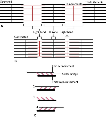
During contraction, the actin and myosin filaments slide over one another and cross-bridges form between the heads of the myosin filaments and the heads of the actin filaments. The cross-bridges swing through an arc, pulling the thin filaments past the thick ones, and the sarcomere shortens. Once this movement is completed the cross-bridge detaches itself from the thin filament and reattaches itself further away – in other words, the cross-bridges between the myosin and actin filaments act as a ratchet mechanism, thus shortening the muscle (Fig. 4.1). This process requires energy input, which is provided by adenosine triphosphate (ATP) molecules; calcium ions are also essential to the process of muscle contraction.
Muscle tone
Anatomy of a muscle
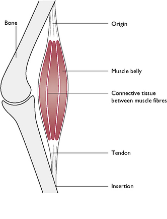
A ‘classically’ shaped muscle (Fig. 4.2) has a thick, fleshy central part called the belly and tapers at each end – the head. Here, the connective tissue muscle sheath is continuous with the dense fibrous connective tissue of the tendon that attaches the muscle to a bone. A muscle is attached to a bone at two points: its starting point is called its origin; this moves least during contraction. The opposite end, where the muscle inserts on the bone, is called the insertion. However, a muscle can have more than one belly, all inserting at one point, in which case the muscle is said to have a number of heads, e.g. the biceps muscle has two heads. The length of the tendon attaching the muscle to a bone will vary, and in some cases tendons are far longer than the muscle itself, e.g. flexor and extensor tendons running over the digits.
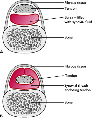
A bursa is a connective tissue sac lined with synovial membrane and filled with synovial fluid. These typically develop between a bony prominence and a tendon, ligament or muscle and their function is to reduce friction between the associated structure and the bone. Sometimes a bursa completely wraps around a tendon forming a synovial or tendon sheath (Fig. 4.3).
The skeletal muscles of the body can be classed as either intrinsic or extrinsic:
The skeletal muscles
To simplify the study of these muscles we can look at one area of the body at a time.
Muscles of the head
Muscles of facial expression
The muscles of facial expression are intrinsic muscles that move the lips, cheeks, nostrils, eyelids and external ears. A number of muscles are responsible for these movements, all of which are innervated by the facial nerve (cranial nerve VII).
Muscles of mastication
The main muscles responsible for the masticatory or ‘chewing’ action of the jaw (Fig. 4.4) are:
Stay updated, free articles. Join our Telegram channel

Full access? Get Clinical Tree


