34 Multiple tooth and jaw fracture
ORAL EXAMINATION – CONSCIOUS
An extremely well-behaved dog that allowed careful examination, which revealed the following:
ORAL EXAMINATION – UNDER GENERAL ANAESTHETIC
In summary, examination under general anaesthesia identified the following:

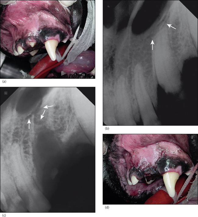
Figure 34.2 Photographs and radiographs centring on 203.
(a) Oblique lateral photograph of the rostral left upper jaw. The complicated crown fracture of 203 is obvious. Note also the wear facet on the distal aspect of 204 due to cage biting.
(b) Rostrocaudal radiograph centring on 203. Note the fracture of the premaxilla level with the apex of 203.
(c) Rostrocaudal radiograph of the extraction socket of 203. The fracture line is even more obvious after extraction of 203.
Stay updated, free articles. Join our Telegram channel

Full access? Get Clinical Tree


