Movement

Chapter 7 Movement


It would be difficult to improve on Liautard’s insistence that the lame horse be examined during movement or his description for how it is best accomplished. Although all parts of the lameness examination are important, the key is the determination of the limb or limbs involved. Not all horses with musculoskeletal problems exhibit lameness that is perceptible under normal conditions, or even by use of high-speed or slow-motion cinematography, gait analysis, or other sophisticated imaging devices. Under most circumstances, however, lameness from pain or a mechanical defect in gait is discernible, and the essence of the lameness examination is to determine the source of the pain. This discussion includes relevant experimental findings to support clinical observations, but sometimes experimental findings are confusing rather than informative.


Gait


Gait, defined as the “manner or style of walking”2 or “the manner of walking or stepping,”3 is used to describe the speed and characteristics of a horse in motion. The natural gaits, those exhibited when a horse is free in a field, are the walk, trot, and gallop.4 The canter is a collected gallop. Other gaits including the pace, running walk, rack (a singlefoot or broken amble), fox trot, and amble are artificial gaits, although some pacers pace “free-legged” (without the use of hobbles) while on the track, either at a slow speed or racing speed, and occasionally a Standardbred (STB) paces free-legged in a field. In some instances a trotter switches from a trot to the pace, but this change usually is exhibited while the horse is performing at speed and may be associated with lameness or interference. Lame trotters usually “make breaks,” going off stride, by switching (breaking) from the trot to the gallop.


The term beat describes the number of foot strikes in a single stride cycle regardless of whether one or more feet strike the ground simultaneously. The following abbreviations are used for limbs: left forelimb (LF), right forelimb (RF), left hindlimb (LH), and right hindlimb (RH). The walk is a four-beat gait in which all four feet strike the ground independently without a period of suspension (in which no feet are on the ground). imageDepending on the part of the stride during which observations begin, the walk can appear to be lateral or diagonal. In general, in a lateral gait, both feet on one side strike the ground before the feet on the contralateral side. In a diagonal gait, one foot strike is followed by a strike of the foot located diagonal and contralateral to the initial foot (e.g., LF followed by the RH).


Lame horses should always be evaluated at the walk.image Stride length should be evaluated and compared with observations at the trot. Stride length and sequence of footfalls are easier to see while horses are walking than while they are trotting. Horses with hindlimb lameness may be examined for failure to track up.5 Horses normally track up, or overtrack. The hind foot is placed in or in front of the imprint of the ipsilateral front foot. Failure to track up usually is caused by hindlimb lameness or poor impulsion, and the hind foot imprint is seen behind that of the ipsilateral front foot.5 Although unusual, occasionally a horse will be observed to pace while walking, a finding that may indicate the presence of neurological disease. In breeds unassociated with the pace or similar gaits, young horses that pace should undergo careful neurological evaluation. Pacing while walking may be completely normal, and in older, “made” horses (horses that have already achieved an upper level of performance) the finding should not be overinterpreted.


Backing is a diagonal, two-beat gait. imageHorses seldom back naturally, but backing commonly is required of horses during performance events, while exiting from a trailer, or while driving. Backing is useful during lameness examination to evaluate certain gait deficits, such as those associated with shivers, stringhalt, and neurological disease. image


The trot is a diagonal, theoretically two-beat gait, and diagonal pairs of limbs move simultaneously. imageThe trot is theoretically a symmetrical gait, meaning both “halves” (beats) of the stride are identical, and at low speed in a sound horse, symmetry is likely achieved. However, at speed, perfect balance and fine management of weight (of the shoes) are necessary for a trotter to be perfectly symmetrical. There is a moment of suspension between impact of each diagonal pair of limbs. Some elite dressage horses do not have a two-beat gait but show advanced diagonal placement. This means that the hindlimb of a diagonal pair of limbs lands first and therefore is the only limb bearing weight. Hindlimb lameness is present in a higher percentage of horses that perform at speed at the trot compared with galloping horses because of differences in weight distribution in the trot and gallop. Compensatory lameness develops in the diagonal paired limb. LF lameness predisposes to RH lameness. Interference between limbs is more common in horses that trot at speed when compared with those that gallop. Likewise, hindlimb lameness is relatively common in dressage horses.


The pace is a symmetrical, lateral, two-beat gait predominantly in STB racehorses and is characterized by movement of lateral pairs of limbs simultaneously (LH and LF; RH and RF), with a moment of suspension between lateral pairs. imagePacers also have a high percentage of hindlimb lameness, but compensatory lameness usually develops in the lateral paired limb. RH lameness predisposes to RF lameness.


The gallop or run is a four-beat gait. In the gallop and the canter the horse leads with the LF or RF, the forelimb that strikes the ground last in the stride sequence. An unrestrained horse usually leads with the LF while turning to the left, or the RF while turning to the right. Fatigue also plays a role. A Thoroughbred (TB) racehorse racing counterclockwise leads with the LF on the turns but immediately after entering the stretch switches to the right lead. Failure to switch leads or constantly switching leads in the gallop or canter may reflect fatigue or lameness.


In a left lead gallop, the RH strikes the ground first, followed in sequence by the LH, RF, and LF, followed by a period of suspension. When a horse is on the right lead, the RF strikes the ground last, propelling the horse into the suspension phase of the stride. It is often assumed that a horse with RF lameness is reluctant to take the right lead. However, bone stress measured in the radius and the third metacarpal bone (McIII) is greater on the nonlead (trailing) forelimb, and thus a lame horse may change leads to protect the nonlead forelimb.6 Ground reaction forces (GRFs) are greater in the trailing (nonlead) forelimb, a fact that supports the clinical observation that horses with forelimb lameness may select leads to protect the lame forelimb. A horse with RF lameness may prefer the right lead, allowing the LF to assume the greater forces and bone stress.5


The canter (lope) is a three-beat gait. In left lead canter the RH strikes the ground first, then the LH and RF land simultaneously, followed by the LF and then a period of suspension. A horse reluctant to take a lead may be trying to compensate for hindlimb lameness. In the right lead the LH must absorb a considerable amount of concussion and then generate propulsive forces. Proneness of this limb to fatigue seems logical, but a consistent change in stride characteristics of fatigued horses to protect the LH was not seen.7 Although the LH strikes the ground first, stance time, flexion of the upper limb joints, and GRF are greater in the RH.5 It could be assumed that a horse lame in the RH would be reluctant to take the right lead and may prefer the left lead.5 Lead and stride characteristics of fatigued and lame horses are complex because of asymmetry of the gait, and forelimb and hindlimb problems could account for failure or reluctance to take a particular lead and inappropriate lead switching.


Young horses early in training or trained horses that are lame may exhibit a disunited canter. The horse may spontaneously change legs behind, but not in front. In changing from left to right lead canter, or vice versa, the forelimbs and hindlimbs should change simultaneously. Horses with back pain or hindlimb lameness may be reluctant to change leads, or may change in front but not behind.


Comparing Lameness Seen at the Walk and Trot


Although lameness score should be determined when trotting a horse (see later), it is useful to compare gait deficits at the walk and trot. In horses with forelimb lameness, an exaggerated head and neck nod may be observed while walking horses with upper limb lameness (genuine shoulder region pain, for instance), and, in fact, head and neck excursion may be more pronounced than that seen while the horse is trotting. Some horses may exhibit odd gait deficits while walking, but deficits may abate at the trot. Horses with rare upper forelimb pain from rib fractures or anomalies may manifest abduction of the forelimb during protraction at the walk but not the trot. imageThe stance phase of the stride is relatively longer at the walk than at the trot. The deep digital flexor tendon and the collateral ligaments of the distal interphalangeal joint are stressed maximally with extension of the distal interphalangeal joint. Thus with severe injuries of either structure lameness may be more severe at the walk than at the trot because of greater extension of the distal interphalangeal joint associated with the relatively long stance duration.5 Horses with hindlimb lameness characterized by a shortened caudal phase of the stride at the walk but shortened cranial phase at the trot often have upper limb lameness, such as that caused by pelvic fractures or osteoarthritis of the coxofemoral joint or from severe pain originating from the foot. In general, horses that have limb flight characteristics that differ between walk and trot should be evaluated carefully because in my experience they often have bona fide pain originating from the upper limb or are affected with an unusual mechanical or neuromuscular deficit.


Relevance of Lameness at a Trot in Hand


Is lameness seen at a trot in hand the same lameness that compromises performance at speed? Is the lameness seen at a trot in hand in a jumping horse the same problem that causes the horse to refuse fences? The answer is usually, but not invariably, yes. For instance, I have seen many STBs show subtle unilateral hindlimb lameness at a trot in hand, but when the horse was later examined at the track and hooked to a cart, pronounced contralateral hindlimb lameness was noted. Differences include the track surface, the act of pulling a cart, the additional weight of the driver, and a faster gait. Lameness often is evaluated on a smooth hard surface useful in exacerbating even subtle problems, but most horses perform on softer surfaces, when other problems may be apparent. More than one lameness problem may exist—one evident at a trot in hand and another while the horse is ridden or driven. Horses can show lameness from one problem when trotted in a straight line but lameness from an entirely different problem while being trotted in a circle. The answer to the first question is complex because performance itself entails the inextricably intertwined relationship between horse and rider or driver and the possible presence of compensatory and coexistent pain. Ideally, to evaluate the role of lameness on poor performance, horses would be evaluated at the speed, at the gait, and in the manner in which they perform, conditions that usually are not always possible to reproduce.


Surface Characteristics and Lameness Examination


The horse should be examined on a smooth, flat surface. I prefer a hard surface, such as pavement or concrete that creates maximal concussion and may exacerbate subtle lameness. However, the clinical relevance of mild lameness seen on hard surfaces, especially on turns, should not be overinterpreted. Many horses that are actively competing successfully show mild lameness on hard surfaces; it is important to understand that the horse does not perform on a surface of pavement, and foot strike patterns and gait could be much improved if the horse performs on firm but forgiving surfaces. Crushed rock, cobblestone, deep sand, or undulating grassy areas and potentially dangerous slippery surfaces should be avoided.


It is important that the surface be nonslip because some horses appear to lack confidence while moving on hard surfaces and alter the gait. In these situations, horses may shorten the stride for protection rather than from lameness.5 Horses with studs or caulks on the shoes may develop induced lameness unrelated to the baseline lameness when trotting on hard surfaces.5


Ideally the gait on hard and soft surfaces should be compared, to help differentiate soft tissue from bony problems. Horses with foot pain usually perform worse on a hard surface. Lameness from soft tissue injuries, such as suspensory desmitis or tendonitis, tends to be worse on soft or deep ground. To evaluate lameness during transition from a hard to a soft surface and vice versa, a horse can be examined while circling on a lunge line in an area where both surfaces coexist side by side.5 Care must be taken to prevent the horse from slipping during this examination.


Determination, Grading, and Characterization of Lameness


Six basic steps are necessary to determine, grade, and characterize lameness. The clinician should determine the following:


The primary or baseline lameness is the gait abnormality before flexion or manipulative tests are used. The practitioner attempts to abolish baseline lameness using analgesic techniques. Lameness in more than one limb may complicate determination of the worst affected limb. It is important to trot a horse even if it is quite lame at a walk, unless an incomplete or stress fracture is suspected. A horse may take a short step with a limb at walk, or can appear very lame, but trot reasonably soundly. Horses with scratches (palmar or plantar pastern dermatitis) or superficial wounds in the palmar or plantar pastern may appear quite lame at walk but trot relatively well. A STB pacer may walk extremely shortly both in front and behind but pace or trot without lameness. However, only the degree of lameness usually differs between a walk and trot. A horse may appear sound at walk and trot in hand, but lameness may be apparent trotting in a circle, in hand or on the lunge, or while being ridden. This lameness now becomes the baseline lameness, and it is under these conditions that the results of diagnostic analgesia should be evaluated. The clinician should try to recognize if the horse has bilateral forelimb or hindlimb lameness that manifests as shortness of stride or poor hindlimb impulsion, or if concurrent forelimb and hindlimb lameness are present. Moderate-to-severe hindlimb lameness can mimic ipsilateral forelimb lameness, although ipsilateral forelimb and hindlimb lameness also occurs. In these horses the veterinarian should perform diagnostic analgesia in the hindlimb first.


Compensatory Lameness


Compensatory (secondary or complementary) lameness results from overloading of the other limbs as a result of a primary lameness. It must be differentiated from the stride-to-stride compensation by a horse to avoid interference injury because of a gait deficit, or lameness, or to shift weight (load) during examination. A compensatory problem develops as the result of predictable compensation a horse may make over time for a primary lameness in a single limb. However, a horse may compensate for lameness in one limb by shortening the stride in another, a stride-to-stride change in gait that is not the result of lameness. For instance, in some trotters with severe LF lameness, reluctance to extend the LF may induce a compensatory shortening of the cranial phase of the stride in the LH limb, creating what appears to be a hike in the LH. If the veterinarian looks only at the hindlimbs, LH lameness may be diagnosed. A trotter performing at speed with LF lameness is likely to develop compensatory lameness in the RF or RH but not in the LH. However, the horse may appear to be hiking (lame) in the LH to avoid interfering with the LF. Elimination of obvious unilateral forelimb lameness usually resolves an ipsilateral pelvic hike. Most horses with pronounced forelimb lameness examined at a trot in hand will have a concomitant shortening of the cranial phase of the stride in the contralateral hindlimb, giving the false impression of coexistent lameness in this limb and vice versa. Experimental results appear to support this clinical impression. In 6 of 10 horses with stance phase forelimb lameness, compensatory movements of horses created a false lameness in the contralateral hindlimb (see following text).8 Once forelimb lameness is abolished using diagnostic analgesia, the shortened cranial phase of the stride in the contralateral hindlimb will abate.


It is often difficult to know which lameness came first, but it is important to understand how horses compensate for lameness and which limbs are at risk to develop compensatory problems. Compensatory problems range from obvious lameness to only mild palpable abnormalities that may still compromise performance. Several predictable patterns of compensatory lameness are possible; the most common is bilateral forelimb or hindlimb lameness. Horses with a specific lameness in one forelimb are at risk to develop the same condition in the opposite forelimb. This tendency may not always be compensation for the primary lameness but may reflect simultaneous injury or degeneration of bone or soft tissue of both limbs. Abnormal loading of forelimbs or hindlimbs, faulty bilateral conformation, and the same shoeing or foot conditions all likely contribute to bilateral, simultaneous lameness. In horses with bilateral lameness, eliminating lameness in one limb usually results in pronounced contralateral limb lameness. Bilateral lameness may affect both limbs equally, resulting in a short, choppy gait. imageThe horse may be lame in one limb while being circled in one direction and lame in the contralateral limb in the opposite direction. image


Racehorses that gallop are most likely to develop compensatory lameness on the contralateral limb or the ipsilateral forelimb or hindlimb. A TB racehorse with a left metatarsophalangeal joint lameness is most likely to develop a similar problem in the RH but may also develop LF lameness. In a trotter the contralateral limb is most at risk, followed by the diagonal forelimb or hindlimb. If a trotter has a right carpal lameness, the left carpus should be examined carefully; compensatory lameness also may occur in the diagonal LH limb. In a pacer the ipsilateral forelimb or hindlimb should be considered after the contralateral limb. In a pacer with LH lameness the RH and LF are at risk.


The most common compensatory lameness is the same problem in the contralateral limb. However, suspensory desmitis is a common compensatory problem in both the contralateral and other limbs. In a TB racehorse or a jumper with LF lameness, RF suspensory desmitis is common. Primary RH lameness may result in suspensory desmitis in the RF. It is logical that soft tissue structures are particularly vulnerable to the effects of overload. Superficial digital flexor tendonitis may develop secondary to a primary problem in the contralateral limb. In trotters a common pattern is primary carpal lameness and compensatory osteoarthritis of the medial femorotibial joint in the diagonal hindlimb, or vice versa.


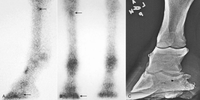
Compensatory lameness also can develop in the same limb. In horses with front foot lameness the suspensory ligament (SL) often is sore, and some horses have suspensory desmitis. In horses with lameness abolished by palmar digital analgesia, most with navicular syndrome, scintigraphic examination revealed increased radiopharmaceutical uptake (IRU) in the proximal palmar aspect of the McIII in 30% of horses, indicating possible abnormal loading of the proximal aspect of the SL (Figure 7-1).9 Complete resolution of lameness may not be achieved until high palmar analgesia is performed.


Horses with primary metatarsophalangeal joint lameness often have associated ipsilateral stifle pain, or vice versa.10 Determination of the primary site of lameness may be difficult without use of diagnostic analgesia and observing that blocking one site abolishes the majority of lameness. This phenomenon may be most common in STBs, but I have recognized it in all types of sport horses and in TB racehorses.


Supporting, Swinging, and Mixed Lameness


Lameness has classically been divided into three categories in an attempt to characterize the motion associated with the lame leg and to assign a cause to the lameness condition. These categories are described and discussed, but I firmly believe that adequate characterization of most lameness conditions is impossible and may be unnecessary.


Supporting limb lameness describes a lameness that results in pain during the weight-bearing phase of the stride. Most lameness conditions are of this type. Supporting limb lameness also has been referred to as stance phase lameness, but this term is inappropriate because the swing phase of the stride is also altered.


Swinging limb lameness describes lameness that primarily affects the way the horse carries the lame limb. However, most horses with painful lameness conditions alter the swing phase of the stride in a typical and repeatable fashion, and it is difficult to make a clear separation between supporting and swinging limb lameness. Swinging limb lameness should be a term reserved for mechanical defects of gait, such as fibrotic myopathy, upward fixation of the patella, stringhalt, or other lameness conditions causing a mechanical restriction of gait. imageIn these horses, lameness is manifested in the swing phase of the stride with no apparent pain. Unfortunately, the term swinging limb lameness often is used inappropriately to describe the gait deficit in horses with painful, supporting limb lameness. Lameness associated with osteochondrosis of the scapulohumeral joint is often described as a swinging limb lameness because of a markedly shortened cranial phase of the stride. Dramatic improvement in the shortened cranial phase of the stride can be achieved by diagnostic analgesia, eliminating pain associated with lameness. Thus the gait deficit is the direct result of pain, and no clear differentiation between supporting and swinging limb lameness can be made. Horses with painful forelimb lameness almost always shorten the cranial phase of the stride, although perhaps not to the extreme as in a horse with authentic scapulohumeral joint lameness. imageHorses with any painful hindlimb lameness consistently shorten the cranial phase of the stride, a reliable clinical indicator of which limb is affected, and when pain is abolished, the cranial phase (swing phase) of the stride improves (lengthens). Because the terminology is confusing and often erroneous, I prefer to avoid use of these terms and simply describe lameness as accurately as possible. For instance, describing a horse as grade 2 of 5 LF lame, with a marked shortening of the cranial phase of the stride reminiscent of other horses I have seen with shoulder region lameness, gives the most accurate and useful information.


There is an erroneous tendency to equate a swinging limb lameness with one that is more evident when the lame limb is on the outside of a circle. Upper limb lameness is often presumed yet not confirmed by diagnostic analgesia. It is logical that if a horse is reluctant to swing a limb forward, the lameness may be most prominent when the lame limb is on the outside of a circle. However, many horses with painful weight-bearing lameness show more pronounced lameness with the limb on the outside of a circle, a finding that neither suggests that lameness originates from the upper limb nor indicates the presence of swinging limb lameness (see following text). The outer limbs must stretch further and cover a larger circumference circle than the inside limbs. Slight temporal differences in the stance and swing phases of the inside and outside limbs are necessary to maintain gait symmetry.5 Therefore the stance phase of the stride may be relatively longer for the inside hindlimb, resulting in extension of the fetlock for a longer time and stress on the suspensory apparatus, whereas for the outside hindlimb covering a longer distance in the same time, there may be greater extension of the fetlock for a relatively short time, but still with stress on the suspensory apparatus. Thus pain associated with hindlimb proximal suspensory desmitis is worse in some horses with the lame or lamer limb on the inside of the circle, whereas with others lameness is accentuated with the lame limb on the outside of a circle.


The results of cinematographic analysis of gait in lame horses seem to support reservation of the term swinging limb lameness for horses with authentic mechanical gait deficits, rather than those induced by painful lameness. In a horse with a supraglenoid tubercle fracture examined at a trot in hand, a marked decrease in the cranial phase of the stride (protraction) was observed, along with a marked head and neck nod. A markedly shortened stride could be equated with swinging leg lameness, but high-speed cinematography showed that the cranial and stance phases of the stride were shorter than in the sound limb.11 A horse with unilateral semitendinosus fibrotic myopathy had a shortened stride length and a shortened cranial phase of the stride, but the stance phase did not differ from that of the unaffected contralateral limb.12


In my experience, most lameness conditions can be considered mixed lameness, with changes in gait during weight bearing or the stance phase and during the swing phase of the stride. With the exception of mechanical defects in gait, I have not been able to categorize the clinical characteristics of most lameness conditions into swinging or supporting limb types. However, it has been suggested that swinging limb lameness is caused by muscle injury; supporting limb lameness is caused by bone, tendon, and ligament injury; and mixed lameness is caused by joint, tendon sheath, and periosteal injury.13 A shortened cranial phase of the stride is a common characteristic in forelimb and hindlimb lameness and should not be considered pathognomonic for the location or type of lameness.


Determining the Location of Lameness


The horse should be observed at both the walk and the trot from the front, behind, and side. I spend most of my time watching the horse move away and then back toward me. Medial-to-lateral limb flight and foot strike can be evaluated only from this perspective, although cranial and caudal aspects of the stride and fetlock drop (see following discussion) can be evaluated only from the side. Most important, evaluation of lameness from this perspective allows the veterinarian to use the horse as a frame of reference. I find it quite useful to evaluate forelimb lameness when the horse is traveling away from me and hindlimb lameness when the horse is traveling toward me. This perspective allows use of the horse’s top line to see a subtle head and neck nod or pelvic hike. Only by observing the horse from the side can the cranial and caudal phases of the stride be determined. When first learning to assess lameness from the side, a linear frame of reference, such as a fence or wall in the background, may be helpful to notice head nod and pelvic hike against an immovable background. Application of pieces of tape or other markers to the horse’s head or a fixed point on the pelvis can assist recognition of upward and downward movement of that body part.


Independent observation of the forelimbs and hindlimbs is needed to understand whether a horse has forelimb or hindlimb lameness or a combination. These observations then are amalgamated to form a final clinical impression.


Recognition of Forelimb Lameness


Forelimb lameness often is easier to recognize than hindlimb lameness. Understanding the concept of the head nod is vital to the correct interpretation of equine lameness. The head and neck elevate or rise when the lame forelimb is bearing weight or hits the ground and nod down or fall when the sound forelimb hits the ground. “When the [forelimb] is the lame one, the movements of the foot and head occur somewhat in unison. When the lame foot is raised, the head is elevated, but only to fall when the sound leg is brought to a rest.”1 Some clinicians find it easier to appreciate the head nod down, whereas others find it easier to recognize elevation of the head.


When slow-motion videotape of lame horses is evaluated, it is immediately obvious that the elevation of the head and neck is much easier to see than the head nod down. In slow motion the horse appears to be elevating the head and neck just before the lame limb hits the ground, and then, during the later portion of the support or stance phase, the head and neck nod down. In fact, in slow motion it is head and neck elevation from a baseline level and later settling of the head and neck (nod downward) that are seen. The head returns or settles to baseline, giving the distinct impression that the horse is unloading the lame limb rather than loading the sound limb. The head and neck nod occurs as the contralateral limb begins the support or stance phase. Both head elevation and falling are present, but head elevation is much easier to detect when it occurs in unison with the lame limb hitting the ground. It is likely that a combination of visual clues allows the clinician to decide the primary forelimb lameness. Quantification of lameness and description of the actions of the lame and compensatory limbs have been attempted using gait analysis systems. In horses with amphotericin-induced carpal lameness, head movements were the most consistent indicator of lameness, followed by sinusoidal motion, or a rising and falling action, of the head and withers.14

Stay updated, free articles. Join our Telegram channel

Jun 4, 2016 | Posted by in EQUINE MEDICINE | Comments Off on Movement

Full access? Get Clinical Tree

Get Clinical Tree app for offline access