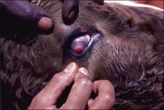
Chapter 29 Moraxella bovis, the cause of infectious bovine keratoconjunctivitis, is the major animal pathogen in the genus. The pathogenicity of M bovis strains depends on the production of type IV pili and a cytotoxin, in addition to other putative virulence factors (Table 29.1). The cytotoxin is a haemolysin and belongs to the repeat-in-toxin family of toxins (Angelos et al. 2001). Strains which are not haemolytic or lack pili are apathogenic for cattle. In addition to the pili, which have a major role in attachment of the organism, filamentous-haemagglutinin-like proteins may be important in the infectivity of the organism (Kakuda et al. 2006). Other enzymes which may play a role in tissue destruction include fibrinolysins, proteases and phospholipases. Acquisition of iron through transferrin and lactoferrin-binding proteins may contribute to virulence also. Table 29.1 Virulence factors of Moraxella bovis The early signs of infectious bovine keratoconjunctivitis are lacrimation, blepharospasm and conjunctivitis. Later an ulcer develops on the cornea. Corneal opacity and oedema surround the ulcer and in severe cases vascularization of the cornea occurs from the limbus to the ulcer. The corneal opacity then involves the entire cornea. In the healing stage, granulation tissue forms on the ulcer floor and a characteristic red cone of granulation tissue will project from the cornea (Fig. 29.1). The granulation tissue and the ulcer itself will eventually regress leaving a white corneal scar. The scar may or may not be permanent. Mild cases are similar, but ulcers resolve without vascularization occurring and most eyes become clinically normal in two to three weeks. Figure 29.1 Young steer with infectious keratoconjunctivitis showing the healing stage with the characteristic red cone of granulation tissue projecting from the cornea.
Moraxella species
Pathogenesis and Pathogenicity
Virulence factor
Encoding gene(s)
Comments
Type IV pili
Pilin gene depends on strain
The genes encoding pilin from the prototype strain of each of the seven serogroups of M. bovis have been characterized by Atwell et al. (1994)
Cytotoxin (MbxA)
mbxA
Member of the RTX family. Haemolytic, cytotoxic and leukotoxic properties (Angelos et al. 2001)
Phospholipase
plb
Displays phospholipase B activity (Farn et al. 2001)
Filamentous haemaglutinin-like protein
flpA and flpB
Genes located on a plasmid, recently described by Kakuda et al. (2006)
Transferrin and lactoferrin-binding proteins
tbpA, tbpB, lbpA and lbpB
Bind bovine transferrin and lactoferrin for use as a source of iron. Characterized by Yu et al. (2002)

![]()
Stay updated, free articles. Join our Telegram channel

Full access? Get Clinical Tree


Moraxella species
Only gold members can continue reading. Log In or Register to continue
