Chapter 16 Miscellaneous Anomalies
Otitis Media/Interna: Calves
ANATOMICAL AND PHYSIOLOGICAL CONSIDERATIONS
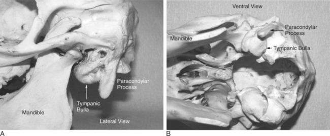
The middle ear comprises the tympanic cavity and auditory tubes lined by mucous membrane. The tympanic cavity, located between the tympanic membrane and internal ear, consists of three parts: the atrium, the epitympanic recess—which contains most of the auditory ossicles—and the large tympanic bulla (Figure 16-1A and B). The function of the middle ear is to transmit sound waves that reach the tympanic membrane through the auditory ossicles to the internal ear. The internal ear consists of two cavities (membranous and osseous labyrinth) in the petrous part of the temporal bone that enclose a complex membranous membrane containing the auditory cells and distal ramification of the auditory nerve. The osseous labyrinth, immediately medial to the tympanic cavity, has three parts: the cochlea, vestibule, and semicircular canals. The membranous labyrinth lies within the osseous labyrinth; it contains supporting cells and hair cells. The distal extremities of the cochlear nerves are located at the base of the hair cells.
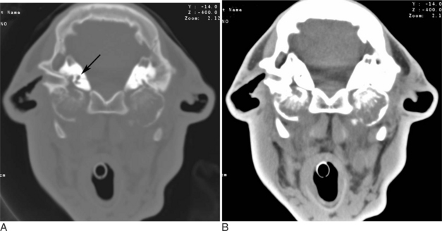
Chronic suppurative otitis media results first from a bacterial infection. The latter may be primary or secondary to a viral infection. This causes inflammation, ulceration, and production of granulation tissue within the middle ear (Figure 16-2B). The cycle of inflammation described above leads to destruction of the bony margins of the middle ear (Figure 16-2A and B).
Stay updated, free articles. Join our Telegram channel

Full access? Get Clinical Tree


