Chapter 6 Minipigs
Introduction
Minipigs are now used in greater numbers in contract research organizations as well as in the pharmaceutical industry. They provide significant advantages over dogs due to their greater tolerance of non-steroidal anti-inflammatory drugs, antihypertensive agents and sympathicomimetic drugs (Dincer 2007). In addition, minipigs are advantageous models for human drugs since their digestion is similar to that of humans, and their smaller size makes them more manageable laboratory animals. The minipig offers many advantages as a model in dermal toxicity because of the similarity between porcine and human skin (Mortensen et al 1998, Lavker et al 1991), including similarities in skin thickness, permeability, pigmentation, allergic reaction and reaction to burning.
In addition, the gastrointestinal system, digestion and metabolism, as well as the renal and immune systems of minipigs are similar to those in man. There are also similarities to humans in reproductive tract histology, physiology, and in cervical and vaginal secretions and vaginal pH (minipigs are used for intravaginal studies) (Jørgensen et al 1998). Finally, cardiovascular physiology and anatomy, ventricular performance, electrophysiology and coronary artery distribution are similar to those in humans.
Cardiovascular system
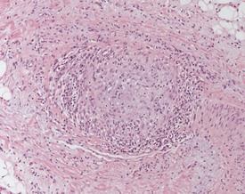
Arteritis and periarteritis are commonly observed in a variety of organs, including the epididymides, heart, intestine, kidney, lung, spleen, stomach and urinary bladder (Fig. 6.1). In the heart, small foci of lymphocytes and occasional areas of myofibre necrosis are observed. Mononuclear inflammatory cells are sometimes encountered within the minipig aorta. Occasionally focal arteritis may be observed in the minipig kidney.
Hemolymphoreticular system
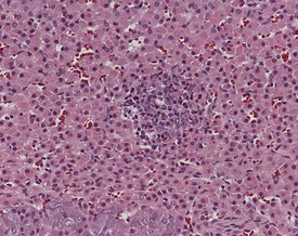
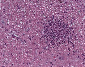
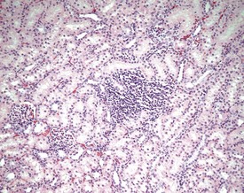
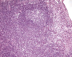
The most common histopathological background lesion noted in minipigs is the focal accumulation of mononuclear inflammatory cells in various organs. These cells are predominantly lymphocytes, macrophages and plasma cells (Madsen et al 1998), and are generally noted in perivascular and interstitial locations (Madsen et al 1998). The foci can be observed in the adrenal glands (Fig. 6.2), cerebrum (Fig. 6.3), epididymis, oesophagus, kidneys (Fig. 6.4), liver, lung, mandibular salivary gland, meninges, parotid gland, rectum, stomach, testes, tongue and vagina (Madsen et al 1998).
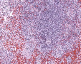
Ellipsoids are pale, eosinophilic structures arranged concentrically around capillaries or small arterioles in the spleen. The ellipsoids display variable prominence; they consist of phagocytic cells and reticular fibres and have been referred to in the literature as Schweigger–Seidel sheaths (Charles 1996) (Fig. 6.5).
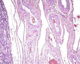
In common with other pig breeds, minipigs display a reversed cortex and medulla in the lymph nodes, i.e. the medulla is present on the outside of the node and the cortex with germinal centres is visible in the middle of the node. In the mesenteric lymph node the presence of a perimesenteric plexus can sometimes be confusing to pathologists (Fig. 6.6). The vascular supply to the lymph nodes differs from that of other mammals. Major nodal arteries subdivide into branches which envelop the node as a capsular network from which further branches penetrate the parenchyma (Charles 1996).
Iron deposits are often visible within macrophages in the sinuses of local lymph nodes (Madsen et al 1998). The widespread iron deposits visible in the minipig are thought to be due to the intramuscular injections of iron administered to neonatal piglets to prevent anaemia (Svendsen et al 1998). In addition, iron deposits can also be encountered in the kidney, liver and mandibular lymph node.
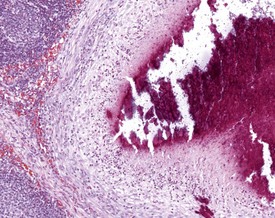
Small lymph abscesses are common in the mandibular node (Dincer 2007) (Fig. 6.7) and are thought to be secondary to local irritation or inflammation. Sinus histiocytosis is also commonly observed in the minipig lymph node. An increase in granulocytic such as eosinophilic cells can sometimes be observed in the sinuses of the mesenteric lymph nodes (Dincer 2007) (Fig. 6.8).
< div class='tao-gold-member'>
Stay updated, free articles. Join our Telegram channel

Full access? Get Clinical Tree


