Methemoglobinemia

Chapter 88 Methemoglobinemia


PATHOPHYSIOLOGY


Oxidation in the Erythrocyte


Reactive species derived from oxygen can cause oxidative damage within the body by transferring or extracting an unpaired electron to or from another molecule. Protective mechanisms that prevent or reverse oxidative damage include proteins that act as free radical scavengers and reducing agents that can remove the unpaired electron from an oxidized molecule.


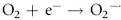
Erythrocytes are especially vulnerable to oxidative damage because they carry oxygen, are exposed to various chemicals in plasma, and have no nucleus or mitochondria.1,3 The lack of cellular organelles renders the membrane the deformability necessary to navigate capillary beds, but results in a cell that is incapable of producing proteins or performing efficient energy production.1 They therefore have a finite number of cell proteins and are reliant on anaerobic respiration to generate energy and reducing agents.1 Oxidants continuously generated in vivo include hydrogen peroxide (H2O2), superoxide free radicals (O2), and hydroxyl radicals (OH·) (Box 88-1).1,3,4 Hemoglobin can undergo autooxidation as an electron is pulled off the hemoglobin onto an oxygen molecule, resulting in the generation of metHb and O2.1,3 Free radicals may also extract electrons by oxidizing deoxyhemoglobin.3 In contrast oxidant toxins can donate an electron to oxyhemoglobin, creating metHb and hydrogen peroxide (Box 88-1).3


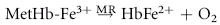
Despite their limited capacity to produce energy and proteins, erythrocytes have many mechanisms to protect themselves from oxidative damage. These include superoxide dismutase, catalase, glutathione peroxidase, glutathione, and metHb reductase (cytochrome b5 reductase) (see Box 88-1).1,3 Glutathione is a tripeptide produced in erythrocytes and composed of glutamic acid, cysteine, and glycine and contains an easily oxidizable sulfhydryl (SH) group.3 It is a powerful antioxidant that operates as a free radical scavenger. Reducing agents such as nicotinamide adenine dinucleotide phosphate (NADPH) and nicotinamide adenine dinucleotide (NADH) are instrumental in reducing oxidized glutathione and metHb back to functional molecules (see Box 88-1).1,3,4


Heinz Bodies


Heinz bodies (HzBs) are aggregates of denatured precipitated hemoglobin within erythrocytes that form as hemoglobin that has undergone oxidative damage is metabolized.1-4 Oxidation of the SH groups of hemoglobin, either through autooxidation, free radical extraction of an electron, or oxidant toxin donation of an electron, causes conformational changes in the globin chains that results in precipitation of the denatured globin.3,4 Aggregates of denatured globin and metabolized metHb clump into HzBs and continue to coalesce until visible, pale structures can be seen within the red blood cell cytoplasm (see Color Plate 88-2, B).4 The complete sequence of events necessary for HzB formation is still being elucidated, but it is thought that formation of metHb is necessary for the development of HzBs.3 Feline hemoglobin is more susceptible to oxidative damage because it has eight SH groups on the globin part of the molecule rather than four, as the canine counterpart does.3,4,7,8


HzBs have an affinity for membrane proteins.4 Binding of a HzB to these proteins causes disruption of anion transport, decreased membrane deformability, and aggregations of membrane protein complexes that may act as autoantibodies.4,7 Numerous HzBs can disrupt the membrane sufficiently to result in “ghost” cells, empty red blood cells with just a cell membrane and HzB remaining, which are associated with oxidation-induced intravascular hemolysis.7 More commonly, however, erythrocytes that have undergone oxidative damage are removed by the mononuclear phagocyte system, particularly within the spleen.7 Rigid cells or cells with large HzBs protruding from the surface will become lodged in the narrow openings between splenic endothelial cells and undergo phagocytosis by the splenic macrophages.3 In most animals, the spleen can perform pitting functions and remove the HzBs from the erythrocyte.3 Feline spleens, however, have an ultrastructural variation and impaired ability to catch and remove oxidized red blood cells.9 As a result of the combination of more SH groups available for oxidation on feline hemoglobin and the unique spleen in this species, healthy cats often have notable HzBs in circulation (with reports up to 96%).9 The reasons that some cats undergo hemolysis with HzB percentages lower than 96% but other cats will have no clinical signs with most of their erythrocytes affected are still unknown.7,9 It is clear, however, that various agents induce oxidative damage in different ways and to varying extents, and the nature of the damage, the amount of affected hemoglobin within a cell, and individual variations seem to determine whether a given cat will develop clinically significant hemolysis.7

Stay updated, free articles. Join our Telegram channel

Sep 10, 2016 | Posted by in SMALL ANIMAL | Comments Off on Methemoglobinemia

Full access? Get Clinical Tree

Get Clinical Tree app for offline access