9 Metazoan/parasitic diseases
Chorioptic mange (leg and tail mange)
Demodectic mange (demodicosis)
Trombiculidiasis (scrub itch/chigger/ harvest mite infestation)
Poultry (red) mite (Dermanyssus gallinae)
Fleas (stickfast or stick-tight fleas)
Culicoides/mosquito irritation
Gasterophilus spp. (bot fly) infestation
Hypodermiasis (warble fly infestation)
Myiasis (calliphorine and screw-worm myiasis)
Pediculosis (louse infestation)
Habronemiasis (summer sores/ bursatti/kunkurs/swamp cancer)
Parasitic/verminous dermatitis
Onchocercal dermatitis (onchocerciasis/microfilariasis)
Oxyuriasis (pinworm) infestation
Chorioptic mange (leg and tail mange)
Profile
This dermatological disease is caused by infestation with Chorioptes equi which primarily affects the distal limb regions but can extend to abdomen, axilla and groin. The parasites are scale-feeders and live on the surface of the skin. Adult mites can survive in bedding and stable floors for some months, especially if there is accumulated skin debris.
The condition is part of the verrucose pastern dermatitis (greasy heel syndrome (see p. 47). The disease is transmitted by direct and indirect contact. Chorioptes equi is possibly species-specific and certainly does not seem to infest in-contact humans, goats, sheep or cattle.
The parasites are easily recognized (see p. 46).
 Key points: Chorioptic mange


1. Common cause of pruritus, worse in housed horses in winter months and commoner in feathered legs. The mites survive well in the stable environment especially where skin debris and dander are present and straw bedding is used. Carrier horses harbour a few mites around the distal limb over summer.
2. Usually many parasites (at varying stages and eggs) but clinical signs can be severe with only a few parasites. Pruritus not always proportional to severity of infestation!
3. Also affects body and neck/head and can be generalized, causing scaling and severe self-trauma. Carrier animals may show no signs.
4. Diagnosis is simple by examining brushings with magnifying glass or microscope
5. Treatment involves repeated anti-parasitic washes, clipping of affected areas and stable hygiene. Important to treat all in-contact horses at the same time. Stable hygiene should be improved. Turn-out generally results in reduced mite numbers.
Clinical signs
Pruritus, irritation and restlessness with foot stamping and biting at legs are prominent early signs. Stabled horses in winter seem more liable to the infestation, with some remission over grazing periods. It is likely that some of these cases remain carriers, perpetuating the infestation within a stable yard. The affected horse seeks every opportunity to rub against or kick on posts, fences, concrete steps, walls and rails. Affected groups of horses can often be heard stamping their feet all night. Irritation is worse in warmer weather. Rubbing of the leg often causes the horse to bite or chew at the site (Fig. 9.1). Rubbing of the tail-base and face can also be encountered.
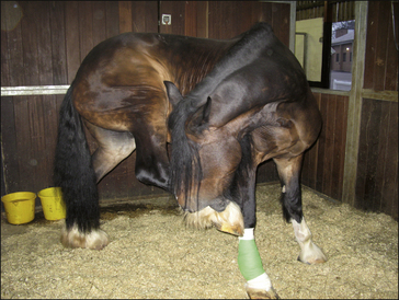
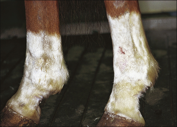
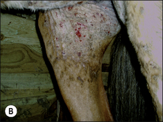
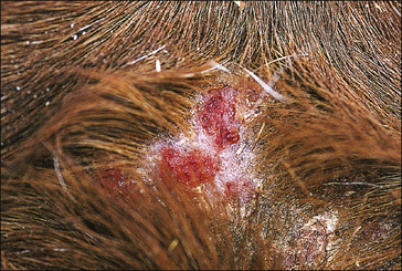
Patchy alopecia with broken hairs (possibly as result of rubbing and biting) and prominent scaling are common signs (Fig. 9.2). Exudation of serum with matting of leg hair, alopecia and scabs may develop over limited or extensive areas. Severe thickening of the skin and secondary bacterial infections arise from the long-standing, persistent self-inflicted trauma and this is one aspect of the verrucose pastern dermatitis syndrome that plagues many heavy draught breeds and the Shire and Clydesdale in particular (Fig. 9.1).
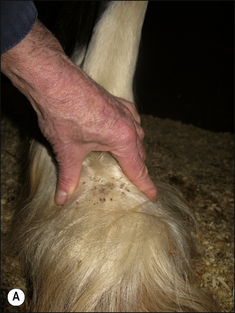
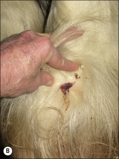
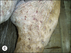
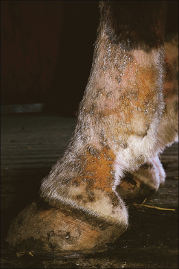
Figure 9.2 Chorioptic mange showing scaling (A) of the pastern region and ulceration (B) which was caused by self-trauma. This horse is the same as that shown in Figure 9.1.
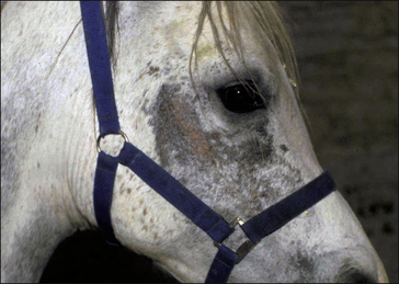
The severity of pruritus is not proportional to the severity of the infestation – some horses will rub a great deal with few mites while others with heavy burdens will show little (or possibly even no) pruritus. The condition is not restricted to horses with feathered limbs (Fig. 9.3).
Repeated episodes of self-inflicted damage can cause progressive thickening of the skin and exudation over the areas concerned. In some cases there may also be a degree of hypersensitivity to the mites. Secondary bacterial and fungal infections, fly strike (myiasis) can combine to produce a true verrucose pastern dermatitis (greasy heel) syndrome (see p. 471).
Differential diagnosis
• Atopy: persistent, variable location; mild to severe pruritus in the absence of any detectable cause in young horses.
• Insect bite hypersensitivity (including Culicoides spp. hypersensitivity/sweet itch): seasonally restricted and usually related to exposure circumstances with severe pruritus of neck and tail and less often ventral midline, head and ears.
• Dermatophilosis (Dermatophilus congolensis): not pruritic and usually obvious inflammatory lesions.
• Dermatophytosis (Trichophyton spp.): only mildly pruritic and characteristic clinical progression and epidemiology.
• Trombiculidiasis (Trombicula autumnalis): mainly affects the limb and head regions; larval mites can be found on brushings or can be seen as orange-coloured moving parasites. Geographically limited to chalky soils.
• Sarcoptic mange (Sarcoptes scabiei): extremely rare condition usually restricted to immunocompromised horses. Extreme whole-body pruritus and mites identifiable in deep skin scrapings.
• Lice infestation (Werneckiella (Damalinia) equi and Haematopinus spp.): obvious pathognomonic macro-parasites identified directly or on brushings.
• Strongyloides westeri and Pelodera strongyloides (parasitic) dermatitis: self-limiting condition affecting young foals, usually on wet/poor pasture; parasites can be identified in washings.
• Cattle tick (nymphal and larval stages): obvious localized visible parasites – not usually pruritic.
• Verrucose pastern dermatitis (greasy heel syndrome): restricted to distal limbs – more often white limbs; can be complicated by secondary parasitic infestations and bacteria.
• Pastern leukocytoclastic vasculitis: restricted, non-pruritic painful condition affecting white limbs only, initially lateral or medial aspects of cannon and pastern.
• Pastern folliculitis: painful, regionally restricted dermatitis.
• Mercurial and other blistering counter-irritants: exposure history, not pruritic.
Diagnostic confirmation
• Mites are easily recognized in groomings and scrapings from the affected regions – the ear margins are a good source of parasites in affected animals (see p. 46). They can usually be easily seen with a magnifying lens on a dark tile or under a low-power microscope.
Treatment
Oral ivermectin paste (at 0.1 mg/kg daily for 7–10 days or 0.2 mg/kg twice at 2-weekly intervals) or moxidectin paste (at 0.4 mg/kg q 14 days) appears to help appreciably. However, this approach does not kill the mites – some do die but others are simply rendered less active and they lay fewer eggs. Therefore this alone does not cure the problem (Littlewood et al 1995). There is no advantage in injecting ivermectin over oral dosing – the intestinal absorption is rapid and provides the same blood concentrations very rapidly. There are significant dangers in injecting the drug into horses apart from its ‘off label’ use. Injectable doramectin show some promise as a therapeutic agent but again the effects are unreliable.
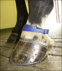
Topical washes of the affected horse and the in-contact animals are the main approach. Affected animals should be clipped out first (although in some cases this is anathema to the owners!). A preliminary warm wash with a selenium sulphide shampoo is a very helpful measure prior to the application of fipronil spray (FrontLine, Merial, UK) (see p. 93) or an anti-parasitic rinse. Application of a cat or dog flea collar impregnated with cypermethrin or permethrin is a very helpful approach (see p. 95) and provides a good measure of protection for some weeks. The collars should be tested on one limb first in case the animal shows a local reaction, and must be fitted with a locking buckle to avoid over-tightening and an elasticized section to prevent the collar ‘snagging’ (Fig. 9.4).
Psoroptic mange
Profile
This is a parasitic skin disease (body mange) primarily of young stabled horses caused by the non-burrowing scale- and fluid-eating mite Psoroptes equi. Psoroptes spp. mites do not appear to be particular about their hosts and so there may be fewer species than once was appreciated. However, it does appear that different feeding habits do occur and so this may be the main species differences. Psoroptes hippotis may be a separate species which may cause some cases of ear irritation and head-shaking. Older horses may become infected if infected younger horses are introduced into the stable. Mites can also be spread by head collars and rugs. Survival in a cool damp environment is possible for up to 2 months or more. Carrier status is not yet established but individual yards are recurrently affected. Degrees of hypersensitivity can occur also.
 Key points: Psoroptic mange


1. Uncommon cause of pruritus largely restricted to the head and ears and tail regions. There may be relatively few parasites.
2. Signs are mainly related to pruritus and localized skin thickening with hyperkeratosis. Severe excoriations can occur and this may be complicated by hypersensitivity reactions.
3. Diagnosis is by identification of the mite in ear debris and skin brushings and scrapings.
4. Treatment options are limited to topical anti-parasitic creams and washes and oral ivermectin or moxidectin. Treatment commonly has to be repeated frequently. Hygiene and avoidance of transfer to other horses is important.
5. It is worth checking other in-contact horses carefully. Once eliminated, the condition is relatively easy to avoid. A few horses become long-term carriers.
Clinical signs
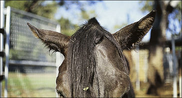
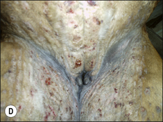
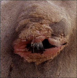
Some affected horses show few or no signs at all but close inspection of the pinnal margin in particular may reveal thickening and hyperkeratosis. Obvious clinical signs attributed to the condition include head-shaking/rubbing and tail-rubbing. Moderate to severe pruritus mostly around the ears, mane, body and tail-head are non-specific but important signs (Fig. 9.5). Pruritus is more severe in warmer weather. Scaling (seborrhoea) and exudation are common, particularly along the margins of the ears along the mane and over the tail-base.
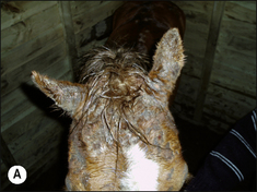
Aural discharge, head-rubbing, head-shaking (with a side-to-side shake) and the ears held flat (‘droopy’ ears) are characteristic of P. hippotis (Fig. 9.6).
Differential diagnosis
• Insect bite hypersensitivity (Culicoides hypersensitivity/sweet itch): seasonally restricted pruritus affecting predominately the mane and tail regions but occasionally the ventral midline and head and ears.
• Chorioptic mange (Chorioptes equi): predominately affects the legs and head regions. Mites easily harvested by brushings.
• Lice (Werneckiella (Damalinia) equi and Haematopinus asini): easily recognized parasites in brushings.
• Oxyuris equi infestation: perineal/perianal pruritus only in unwormed horses.
• Stickfast flea, Tyroglyphus spp. mites: perineal and ear inflammation and irritation.
• Spinose ear tick (Otobius megnini), cattle tick larvae, other ticks: head-shaking, easily viewed auriscopically.
• Atopy (feed or inhaled allergy): non-seasonal, variable over body, onset in young horse.
• Conchal cyst: non-pruritic single discharging sinus tract on pinna or below.
• Idiopathic or photic head-shaking: characteristic neurological disease.
• Aural haematoma: very rare secondary event to shaking and rubbing.
• Over-strength insecticides: history of application – discomfort and inflammation localized, not pruritic.
Diagnostic confirmation
• Samples of ear wax or skin groomings can be examined with a hand lens on a dark tile – small white moving dots will be seen.
• Confirmation by microscopic identification of the mites (see p. 45).
Sarcoptic mange
Clinical signs
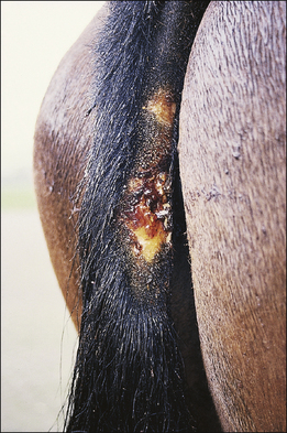
This condition is characterized by intense, almost uncontrollable pruritus. Usually it is reported to start in one site but then rapidly spread to involve the whole body. Lesions frequently begin on the head, neck and ears and may then spread over the entire body (Fig. 9.7).
 Key points: Sarcoptic mange


1. A very rare skin disease causing severe or extreme pruritus. Most affected horses are immunocompromised. Contact with affected pigs, small ruminants and humans can be a factor. Pruritus is a result of dermal burrowing and hypersensitivity responses.
2. Pruritus is usually severe, especially in compromised cases where generalized excoriations and severe and distressing itching occur. Severe damage and secondary skin infections are likely. Pruritus is worse if the skin is warmed by sunlight or by rugging.
3. Parasites can be identified in skin scrapings (seldom in brushings). Multiple skin scrapings will maximize the chance of finding the parasite, which is easily recognized. Biopsy may be lucky – a parasite or its track may be recognizable in the generally inflamed skin; it is often complicated by inflammation from self-trauma.
4. It is essential to address any underlying immunocompromising disorder at the outset. Treatment involves repeated anti-parasitic washes (lindane or malathion), clipping of affected areas and stable hygiene. Oral ivermectin or moxidectin may help and ‘off label’ doramectin injections can be justified. The source of the infestation should be sought.
5. The prognosis is poor and complicated by any underlying disease.
Differential diagnosis
• Insect bite hypersensitivity (Culicoides spp. hypersensitivity/sweet itch) or irritation from swarm insect attacks: characteristic regional pruritus occurring in seasonal patterns year on year; transient and better when avoiding possible contact with cause.
• Black-fly worry (Simulium spp.): bites are obvious and localized to head.
• Chorioptic mange (Chorioptes equi): localized, mild to moderate pruritus usually restricted to distal limbs and tail and head; parasites easily identified.
• Forage/scrub itch/chiggers/harvest mite irritation (Trombicula spp.): geographically limited, regional pruritus on legs and face. Parasites can be seen.
• Onchocerciasis (Onchocerca cervicalis) (particularly following ivermectin anthelmintic administration): localized mild pruritus in defined areas; geographically restricted.
• Larval cattle tick infestation: not pruritic – worry rather than pruritus, parasites obvious.
• Atopy (feed and inhalant allergy): young horse onset of mild to severe pruritus.
Diagnostic confirmation
• Multiple deep skin scrapings are usually required as there are often few parasites.
• Microscopic identification of Sarcoptes scabiei var. equi (see p. 44). A negative skin scraping may not be significant as the parasite is sometimes very difficult to locate.
• Biopsy of affected regions may reveal parasites or tracts but this is often complicated by inflammatory changes from self-trauma.
Demodectic mange (demodicosis)
Clinical signs
The infestation is invariably asymptomatic in normal horses. Lowering of general body immunity by other conditions (such as debility, concurrent disease, stress or pituitary adenoma) may influence the likelihood of clinical signs developing.
 Key points: Demodectic mange


1. A rare skin infestation causing few symptoms. Demodex spp. are probably best regarded as a normal commensal (and opportunistic pathogen) of the skin of large animals. They may become pathogenic in immunocompromised horses.
2. Most cases are identified incidentally.
3. Parasites can be identified in skin scrapings (not in brushings) taken from the eyelids in particular. Multiple skin scrapings will maximize the chance of finding the parasite, which is very easily recognized. The parasite may be recognizable incidentally in biopsies taken for other reasons.
4. Treatment is not usually necessary – the role of regular avermectin anthelmintic doses is not established but this may reduce the overall prevalence still further.
5. The disease is incidental and seldom if ever significant except as part of an immunocompromise condition (stress, debility, steroid or lymphosarcoma).
Patchy alopecia sometimes with scaling, papules and pustules particularly over the face, neck, shoulders and forelimbs may be associated with the condition and/or the underlying immunocompromise. Some cases of leukotrichia and (possibly) leukoderma around the eyes may be caused by this parasite (Fig. 9.8).
Diagnostic confirmation
• Skin scrapings and biopsy are the only way to identify it. It is best to squeeze the skin firmly during the scraping procedure, which should continue until blood is drawn. The presence of large numbers of mites is suggestive of clinical disease while a single mite may not be significant.
Trombiculidiasis (scrub itch/chigger/harvest mite infestation)
Profile
A second group of mites behaving in a similar way are the so-called forage mites. These are usually larger and heavier and often have a more hairy appearance. They inhabit straw bedding and so clinical signs are restricted to housed horses. The individual behaviour of these opportunistic parasites varies and so the patterns of disease associated with them are unpredictable. Some are surface feeders and others are simply irritants as they move around in the hair coat (usually of the distal limbs).
 Key points: Trombiculidiasis


1. A common, usually transient, seasonal pruritic skin infestation of pastured horses. Regional differences occur in the species involved and the circumstances but most are restricted to chalky soils. Forage mites tend to affect stabled horses − especially those on straw bedding.
2. Mild to severe regionally restricted pruritus under defined management conditions. Usually affects the head and legs more severely but can involve the whole body. Changes in management usually improve matters.
3. LarvalTrombicula spp. parasites can be identified directly as yellow-red or orange dots on the skin. Forage mite species are much larger and usually have a hairy appearance.
4. Treatment is helpful – permethrin washes or fipronil sprays are helpful. Cat flea collars around the legs can help also. The value of avermectin anthelmintic doses is not established but they may reduce the overall prevalence.
5. The disease is incidental and only rarely significant except as part of an immunocompromise condition (stress, debility, steroid or lymphosarcoma).
Tyroglyphus spp. mites occur only in grain and can incidentally affect horses.
Clinical signs
Individual bites are papular, inflamed and exudative and may coalesce to give much wider areas of exudative dermatitis. The condition usually affects the head (nose and face) and the lower legs in late summer and autumn (Fig. 9.9). Repeated and severe biting at the lower legs results in small or larger hairless areas which may become obviously inflamed and exudative after 2–3 days (Fig. 9.10). Irritability, leg-stamping, nose-rubbing and head-shaking reflect the intensely pruritic nature of the infestation – some of the response may reflect a hypersensitivity. Small papules and wheals develop on distal limbs, nose, neck (Fig. CD9 • 1A–C) and the ventral abdomen (where it can cause a form of ventral midline dermatitis). Exudation and superficial inflammation reflect both the local reactions to bites and the self-inflicted trauma. In some circumstances the whole body can be affected.
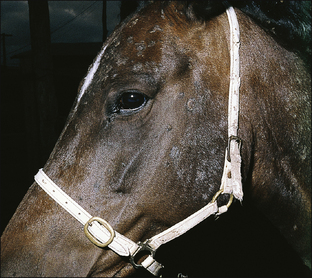
Figure 9.9 Trombiculidiasis. Diffuse dermatitis of the face caused by Trombicula autumnalis infestation.
Differential diagnosis
• Poultry (red) mite (Dermanyssus gallinae) infestation: recognizable parasite, and chicken/bird contact is usual.
• Stomoxys calcitrans spp. irritation and bites: individual bites usually on the body and no residual attached parasite. The bite focus is usually dark purple in colour and very obvious.
• Onchocercal (microfilarial) dermatitis (Onchocerca cervicalis): chronic skin thickening and mildly pruritic. Geographically restricted.
• Chorioptic/Psoroptic mange (Chorioptes equi, Psoroptes equi/hippotis): distinctive parasites detected – may be present in addition.
• Tick larvae infestation: attached larger parasites; seldom pruritic.
• Lice (Werneckiella (Damalinia) equi, Haematopinus asini): large parasites affecting the body trunk and neck mainly.
• Plant irritation (e.g. thistles): exposure related, transient, individuals involved – self-resolving.
• Contact sensitivity/allergy: exposure related, individual horse involved.
Diagnostic confirmation
• Skin brushings are diagnostic – sometimes several different parasites are identified.
• As the mites fall off the host after feeding they are sometimes difficult to find, but aggregations of the red-orange mites feeding at one site make it easier to find on some cases. Individual lesions may be recognizable under a magnifying glass as a papule with a bright red/orange parasite in the centre.
• Extensive examination of groomings may be needed at an early stage in the onset of clinical signs. The use of a dark tile is helpful to show up the orange-red nymphs, although some can be almost colourless (see p. 47).
• Forage mites are very obvious, being much larger and fatter.
Poultry (red) mite (Dermanyssus gallinae)
Profile
 Key points: Poultry (red) mite


1. A pruritic condition due to incidental/opportunistic bites of a variety of poultry blood-feeding ectoparasites including (most often)Dermanyssus gallinae Poultry or pigeon exposure is always involved.
2. Pruritus restricted to specific regions such as the back, face and legs is the usual presentation. Some individuals will be more/less affected than others.
3. The parasites are easily identified but they do not stay on the horse for long. Therefore extensive and repeated brushings are often required. Circumstantial evidence is often strong! The mites may be found in and on wooden rails used for perching.
4. Treatment is not usually necessary – but washes and dusting with a permethrin compound will help limit the effects. Washing any wooden roosting/nesting sites and eliminating or treating the chickens is also helpful.
5. Removal of the contact between the horses and the poultry is obligatory and curative but removal of the poultry can in fact make matters worse (at least in the short term) unless the environment is cleaned as well.
Clinical signs
There is severe, often progressive, pruritus over the face and distal limbs initially but often extending to the whole body. Horses become irritable and stamp and bite at the body and legs (Fig. 9.11). Some cases cause severe excoriation – the extent of pruritus may be disproportionate to the number of mites present. Small pruritic skin papules and crusts may be present in areas readily accessible to the mite (legs, face and ventrum).
Differential diagnosis
• Lice (Werneckiella (Damalinia) equi, Haematopinus asini): characteristic parasites easily identified in brushings.
• Chorioptic, sarcoptic or psoroptic mange (Chorioptes equi, Sarcoptes scabiei, Psoroptes equi/hippotis): characteristic parasites recognized – may coexist.
• Insect bite hypersensitivity (Culicoides hypersensitivity/sweet itch) and irritation (ventral biting species in particular).
• Stomoxys calcitrans fly bites/irritation: single or few and bite sites are more severe and isolated.
• Forage/’scrub itch’, harvest (Trombicula spp.) mites: restricted areas affected and mites characteristic on visual inspection and brushings.
• Larval tick infestation: usually miliary tiny larval or nymphal ticks present; irritational rather than pruritic but can be severe. Recognizable parasites on brushings.
• Larval nematode dermatitis (Strongyloides westeri).
• Food allergy/atopy: rare, persistent, mild-to-moderate shifting pruritus in young horse without any detectable cause.
Diagnostic confirmation
• Poultry contact is always involved. The mites and their eggs can be found in bedding and crevices in walls and on wooden poles and poultry perches (see p. 48).
• Groomings show small, mobile, red nymphal mites which are very visible on a black tile. Microscopic identification of mites.
Fleas (stickfast or stick-tight fleas)
Profile
 Key points: Fleas


1. Fleas from farm stock and domestic pets as well as wild animals can affect horses because they are not host-specific. The poultry flea can also infest horses but prefers its own host. Sporadic outbreaks can occur but once established the fleas are extremely difficult to eliminate. Fleas remain attached to the skin for some hours and so they can sometimes be seen.
2. The signs are seldom obvious but localized foci of mild to intense pruritus can be identified usually after the flea has left the site. Examination of the animals and birds in contact is helpful. Sometimes the humans will also be bitten and this is strong epidemiological evidence.
3. Diagnosis can be made by identifying and capturing the fleas. A small bleb of petroleum jelly will prevent them from escaping! Flea dirt can be found by combing the hair in the affected regions. The dander is placed on damp blotting paper and observed for red discolouring of the blotting paper as the ‘flea dirt’ dissolves.
4. Anti-parasitic washes may be effective but their effects are short and so they may have to be repeated often.
5. Control is the solution – either a concerted effort to treat the animals and poultry and the environment together using controlling sprays such as imidocloprid or separation of the source animals/birds from the horses are the main options.
Differential diagnosis
• Insect bite hypersensitivity (Culicoides spp. hypersensitivity/sweet itch) or irritation from swarm attack: characteristic relationship to outdoor living in summer months; localized to neck and tail-head or ventrum (or both).
• Chorioptic, psoroptic and other mange mites: leg and head pruritus with characteristic patterns of itch and detectable mites.
• Lice (Werneckiella (Damalinia) equi, Haematopinus asini): visible lice and eggs. Generalized pruritus and moth-eaten hair coat.
• Other ectoparasites, e.g. spinose ear tick (Otobius megnini): usually identifiable.
Treatment
The fleas are very hardy and so elimination can be a real problem. Sustained treatment of source animals/birds and their own environments or significant and strict separation of horses from source animals including poultry can be effective. Often, however, removal of the others species simply results in a bigger challenge to the remaining animals. Ongoing control measures including regular stable spraying with anti-flea aerosols such as imidocloprid may be helpful but there is no reported information on the likely benefits. Poultry farmers and advice organizations may be a good source of information on the latest effective control strategies.
Culicoides/mosquito irritation
Profile
The Culicoides family (also known as ‘no-see-ums’, midges or gnats) includes many hundreds of species each with its own geographical location and breeding/feeding habits and some have a host preference. Many geographically restricted species of mosquito and Culicoides attack horses. Individual species of Culicoides tend to attack different areas of the horse (Pascoe 1973). It is important epidemiologically and clinically to know what Culicoides spp. midges there are in any particular geographical region.
Apart from their nuisance effects both Culicoides spp. and mosquitoes (Culex spp., Aedes spp. and Anopheles spp.) have an important second role in the transmission of some significant viral diseases (including the viral encephalitides such as West Nile virus and African horse sickness) and some other infectious diseases (including those caused by Onchocerca cervicalis and Leishmania spp.). These transmissions often require specific species of Culicoides; for example African horse sickness is probably transmitted only by Culicoides imicola. Both Culicoides and mosquitoes are also involved in the hypersensitivity disorders known as insect bite hypersensitivity (sweet itch/Queensland itch) (see p. 284) (Fadok & Greiner 1990).
 Key points: Culicoides /mosquito irritation


1. This is a worldwide problem relating to the bites of Culicoides and other mosquito species. The condition results in a painful, papular and pruritic skin disorder and in some cases worry/behavioural changes. Swarm attacks are common but some individuals seem able to tolerate the multitudes of bites without any problem. The greater importance lies in the transmission of important diseases and to individuals that develop a hypersensitivity to the bites.
2. Swarm attacks cause more problems because of the large number of bites. Bites are restricted to the preferred feeding sites of the prevalent species. Individual bites are papular, pruritic and sometimes oedematous. Large numbers of bites in one location cause coalescent areas of erythema, pain and pruritus.
3. Diagnosis is usually simple. Known circumstances of exposure are almost impossible to avoid.
4. Avoidance by stabling or insect repellents can be effective to some extent. Severe attacks may warrant short-term systemic corticosteroid treatment. Localized bite sites may benefit from topical corticosteroids/antihistamines and local anaesthetic creams.
5. A relatively few cases develop progressive hypersensitivity problems (insect bite hypersensitivity). Eliminating populations of Culicoides is probably impossible but controlling numbers through traps, breeding site disruption and environmental management can help.
Clinical signs
 .
.Secondary dermal bacterial or fungal infections can occur if self-trauma is significant.
In contrast to the allergic disorder which affects individuals, most or all of the horses in a group will usually be affected to some extent simply as a result of common exposure. A few individuals may not be affected in spite of the challenge of many bites. Self-inflicted trauma can cause moderate to severe superficial skin abrasions and excoriation with serum exudate (Fig. 9.12).
Individual horses may develop the allergic condition even after a single attack, thereafter remaining allergic and becoming progressively more affected in succeeding seasons. Some breeds are apparently more sensitive and others (such as the Icelandic and Welsh ponies and the Shire horse) are more inclined to develop an allergic reaction.
The relationship between insect bites (of any type) and the common nodular skin disease ‘collagen necrobiosis’ or ‘collagenolytic granuloma’ (see p. 292) is not established. Some cases of this disorder have obvious tracts leading to the skin while others do not. The location of the nodules on the dorsum of the back and the sides of the neck tends to indicate that mosquito and Culicoides bites are at least less likely to be involved than other heavier biting insects.
Stay updated, free articles. Join our Telegram channel

Full access? Get Clinical Tree


