Metabolic Diseases

Chapter 14 Metabolic Diseases


The common metabolic problems of early lactation, milk fever and ketosis, are really management diseases. At the herd level, disease does or does not occur as a function of how cows are fed and handled during the late dry period and during transition to the nutrient-dense rations needed to support high milk production in early lactation. Because infectious diseases are more effectively controlled by sound immunization, the economic importance of these common metabolic disorders and their prevention by sound nutritional and herd management has assumed ever greater relevance on the modern dairy. Feeding management includes sources, storage, preparation, ration formulation, delivery, and access. Good feeding management must be coupled with providing an environment as comfortable as possible to facilitate maximal feed consumption. In investigating herd problems of excessive metabolic diseases, all these factors must be considered. Individual cows may be predisposed to metabolic problems as a result of improper body conditioning, concurrent illness, genetics, and any other events that may decrease dry matter intake. In addition to calcium, the other macrominerals of relevance in dairy cattle are potassium, magnesium, and phosphorous, and although disorders involving these elements are of far lesser importance than hypocalcemia, they will also be considered within this chapter.


KETOSIS: CAUSES, CLASSIFICATION, AND PATHOPHYSIOLOGY


Ketosis occurs when cows are in negative energy balance. This most commonly happens in the last 2 weeks of pregnancy or in early lactation. In the last weeks of gestation hormonal factors and decreased rumen capacity may cause a decrease in nutrient intake and/or an increase in lipolysis. At parturition the major demand is that of milk production such that negative energy balance continues. Although the volume of milk production and lactose formation is the predominant demand for energy, there is also a secondary (or possibly primary in some cows) lipid demand for milk fat synthesis. It appears obvious to us that our ability to feed cows in the 2 weeks before freshening to 4 weeks after calving has not kept up with our advancements in genetics for milk production. There are many categories of ketosis in cattle but most involve a similar pathophysiology of lipolysis, excessive release of nonesterified free fatty acids (NEFAs), inadequate hepatic metabolism of increased amounts of NEFAs (incomplete oxidation results in production of ketone bodies), increased fatty acid storage as triacylglycerols in the liver (kidney and muscle to a lesser extent), and, in some cows, decreased hepatic secretion of very low-density lipids (VLDLs). Certain cows with primary ketosis may be genetically predisposed to hepatic lipidosis because of their inability to properly remove triglycerides from the liver.


Pregnancy toxemia is mostly related to an inability to meet energy requirements for fetal development. It is equally common in heifers as multiparous cows and may be predisposed to by twin fetuses. Transient secondary ketosis can be defined as a transient increase in plasma beta-hydroxybutyrate (BHB) caused by a decline in feed intake directly related to another disorder (e.g., left displacement of the abomasum [LDA]).


Subclinical ketosis refers to “clinically normal” cows in the first weeks of lactation that have BHB values greater than 1400 μmol/L or 14.4 mg/dl. Clinical effects can be seen as excessive weight loss, decreased appetite and production, and diminished reproductive performance. Subclinical ketosis may be present in 30% to 50% of early lactation cows in some herds.


Primary clinical ketosis will refer to ketosis in early lactation cows (usually between 1 and 3 weeks in milk, and most commonly in cows in their second to fourth lactation) that are seemingly well fed, in proper body condition before calving, and have no other medical illness. These cows often have BHB levels greater than 3000 μmol/L or 26 mg/dl. Fat cow/fatty liver syndrome refers to the overly conditioned cow that becomes ill just before or at parturition and suffers from marked anorexia, relapsing milk fever, retained placenta, myopathy, and sepsis.


Hepatic lipidosis may take at least three forms: (1) clinically silent in subclinical ketosis, (2) chronic fat mobilization following early-onset periparturient ketosis with an individual susceptibility as a result of either genetics and/or periparturient overconditioning, and (3) periparturient ketosis in the obese cow with massive lipid accumulation in the liver within the first days of lactation.


Clinical Signs and Diagnosis of Ketosis


Primary or spontaneous ketosis is most common in the first month of lactation, with the majority of cases occurring between 2 and 4 weeks of lactation. Cows with either ketosis early (first week) in lactation or cows with persistent ketosis beyond 4 weeks of lactation are most likely to have more marked hepatic lipidosis. Cows with primary ketosis have reduced feed intake of total mixed rations (TMRs) and may prefer forages over concentrates if ingredient fed. Temperature, pulse, and respiration are normal or occasionally subnormal. The rumen in TMR-fed cows will be reduced in volume, have a lower contraction frequency, and also typically have a small fiber mat. In ingredient-fed cows, the rumen may be normal in size but with a large, doughy fiber mat. It is common to hear the heartbeat while listening to the rumen of affected cows. Ketones may be detected in the breath, urine, or milk. Some sensitive individuals can easily recognize this odor. A urine test for acetoacetate is widely available and is the most sensitive test, although specificity is not as high as with milk ketone tests. A color change to purple indicates the presence of acetoacetate (Figure 14-1). The rate and intensity of change are indicative of acetoacetate concentration, but the urine acetoacetate test may be affected by the hydration status of the cow and the concentration of the urine. Many cows with primary ketosis give a strong purple color on the urine test, although the urine of individuals with hepatic lipidosis may only cause a lighter purple coloration. The manure is drier in consistency than herdmates at the same stage of lactation. Affected cows appear dull with a dry hair coat and piloerection. Neurological signs such as persistent licking at herself or objects, aggressive behavior, and unusual head carriage may be seen with nervous ketosis. The pathogenesis of nervous ketosis is unknown. Inability to rise or ataxia resulting from weakness may be seen in some cows with primary ketosis, and these signs are directly related to hypoglycemia. Metabolic acidosis may occur in some cows and, although unpredictable, can be severe (bicarbonate of as low as 12 mEq/L) in a few cows.


Cows with secondary ketosis have clinical signs related to the primary disease (most often displaced abomasum). Except for metritis, ketosis is rare in cows with systemic disorders such as peritonitis, septic mastitis, and salmonellosis. The urine ketostrips will often be a light purple color with secondary ketosis but may be dark purple if the cow is dehydrated and the urine concentrated. Therapy should correct the primary problem, and the ketosis should then resolve. If the ketosis persists, primary ketosis may be present. A proportion of cows with abomasal displacements will have primary ketosis, which is not surprising because there is a proven association between the two disorders. If BHB is measured and gives a concentration of greater than 1400 μmol/L, this may indicate primary ketosis.


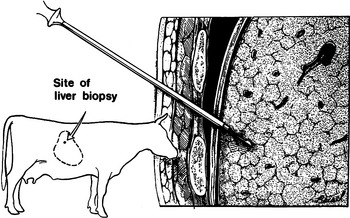
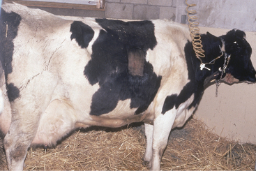
Cows with persistent ketosis for 1 to 7 weeks usually have hepatic lipidosis. Ultrasound examination or biopsy of the liver (Figure 14-2) can be used to confirm hepatic lipidosis, but this is seldom required because the diagnosis is easy but treatment more difficult. Cows with chronic ketosis/fat mobilization and hepatic lipidosis lose considerable amounts of weight, have a poor appetite, but continue to produce moderate amounts of milk considering their poor feed intake (Figure 14-3). Affected cows may appear weak, which could be caused by hypoglycemia, muscle weakness from fatty accumulation in muscle, and/or hypokalemia. Some cows may die, be sold, or have complications caused by frequent treatment (e.g., phlebitis from glucose administration, oral trauma from forced feeding). Serum concentrations of hepatic-derived enzymes (aspartate aminotransferase [AST], gamma glutamyl transferase [GGT], and sorbitol dehydrogenase [SDH]) are often elevated, and serum cholesterol is frequently low in cows with hepatic lipidosis. However, these values are not consistently abnormal. Serum cholesterol generally returns toward normal value as the cow begins to eat better.


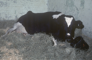
Cows that are overconditioned before parturition and have periparturient ketosis (although a urine ketone test may be only weakly positive) rapidly develop hepatic lipidosis and have life-threatening illness (Figure 14-4). These cows have recurrent hypocalcemia and recumbency and, because of their heavy weight, often develop fatal myopathy (Figure 14-5). Most of these obese/periparturient ketosis/hepatic lipidosis cows have retained placenta and may die of septic metritis even without a fetid smelling discharge (Figure 14-6). Their predisposition to sepsis with mild to moderate metritis may be caused by excessive fat deposition in the liver and diminished hepatic macrophage (Kupffer cells) function. Affected cows may also develop septic mastitis with repeated episodes of recumbency.
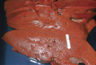


Cows in late pregnancy may become ketotic. This usually occurs with multiple fetuses and is triggered by some other illness or external event that restricts access to feed. Early signs are identical to lactational ketosis. Without prompt treatment, the signs progress to extreme constipation followed by recumbency, renal failure, and death. Cows do not become blind as do sheep with pregnancy toxemia.


Treatment


Treatment for ketosis is aimed at restoring energy metabolism to normal for milk production. The three most commonly used treatments are 500 ml of 50% dextrose given intravenously (IV) once or twice, glucocorticoid administration (e.g., 10 to 20 mg of dexamethasone once), and 300 ml of propylene glycol orally once or twice a day for 5 days. These treatments may be combined to suit the needs of the case and the abilities of the herdsman. The propylene glycol should be given as a drench and not mixed in the feed. Full recovery requires the return to normal feed intake, and supportive therapy may need to be continued for several days to allow time for the cow to maintain normoglycemia. Offering a choice of feedstuffs (i.e., brewer’s yeast) may help in restoring the cow’s appetite. Cows with nervous ketosis can be treated with chloral hydrate (40 g orally daily), which serves as both a sedative and as a substrate for glucogenic-producing bacteria.


Cows with ketosis of pregnancy require more rapid intervention to prevent irreversible hepatic lipidosis and multiorgan failure. Induction of parturition or surgical delivery of the calves may be required. Intensive support of the cow with dextrose and force feeding is necessary. If therapy is discontinued in the first few days after parturition, these cows often have serious, sometimes fatal, relapses of ketosis within 48 hours.


Cows with ketosis and hepatic lipidosis or “fatty liver disease” are challenging cases to treat. Cows with chronic fat mobilization and ketosis/hepatic lipidosis are often the “best cow in the herd” and produce a high milk volume. These cows do not get better overnight with any treatment and in fact may have already been treated with the above listed traditional therapy for ketosis for 1 to 3 weeks before veterinary attention is sought. Treatment should include continual 5% glucose administration in balanced electrolyte solutions with 40 mEq of KCl added per liter of fluid. Insulin (200 IU of zinc protamine, which can be purchased from compounding pharmacies) should be given subcutaneously (SQ) every 24 to 36 hours if a continuous glucose infusion is used. Insulin will promote glucose uptake in peripheral sites, which should inhibit lipolysis. Interestingly the mammary gland and brain of the dairy cow do not require insulin for glucose uptake. Another method of increasing insulin concentration is to give 250-ml boluses of glucose IV twice daily. An attempt should be made to prevent persistent hyperglycemia because this will cause excessive fluid loss in the urine and the hyperglycemia/hyperinsulinemia may predispose to abomasal displacements. Niacin (12 g orally daily) will also inhibit lipolysis and is frequently administered daily to cows with chronic ketosis. Multi-B vitamins are commonly administered (slowly IV) on a daily basis. The most important treatment of cows with chronic fat mobilization and hepatic lipidosis is twice-daily forced feeding. Alfalfa meal, 4 oz of KCl, and rumen transfaunation from a healthy donor cow is our traditional gruel. If these treatments do not appear to be effective after 3 to 5 days, then it may be necessary to reduce the cows’ milk production by milking for 1 minute twice daily until the negative energy balance cycle is broken. Cows should test negative on the California mastitis test to qualify for the controlled milking. Usually the limited milking is required for 4 to 7 days before the ketosis is permanently resolved. We have performed this on many cows with chronic fat mobilization, and it, along with previously mentioned treatments, has been successful in all but one case. Additionally, owners have reported the milk production for the remainder of the lactation was very good. Although cows with chronic fat mobilization have delayed time of estrus and their production is diminished during the first 6 weeks of lactation, their prognosis for complete recovery is excellent. Time to recovery is variable, but most cows are well by 6 to 8 weeks into lactation. The most frequent complication associated with treatment of these cows is thrombophlebitis caused by multiple IV administrations of dextrose.


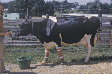
Treatment of periparturient overweight cows with ketosis and hepatic lipidosis is intensive. Affected cows are administered IV fluids to combat hypotension and lactic acidosis. Glucose and calcium are often added to the fluids, although baseline blood glucose levels may be high in some of the cows. The cows should be force fed as described above and have only limited milk removed (if there is mastitis in a quarter, it should be stripped and intramammary antibiotics administered). Insulin therapy can be used as described previously for cows with chronic fat mobilization. Reduced neutrophil and hepatic macrophage function in these cows may allow septic conditions such as even mild metritis or mastitis to overwhelm the patient. Fresh feed, clean water, and salt should be available, and the cow should be housed in either a large well-bedded box stall with excellent footing or in a grass paddock. Along with sepsis, musculoskeletal injury is the most common reason for euthanasia of overweight cows with periparturient hepatic lipidosis. Every effort should be made to maintain calcium levels within normal limits by either slow continuous infusion or SQ administration; ideally ionized calcium should be closely monitored and the cow housed in an area that will provide the best comfort for standing up and lying down. If there has been any difficulty in rising, the cow should be administered flunixin meglumine (500 mg once or even twice daily if needed). The knees should be wrapped with soft cotton bandages to provide protection to the carpus area, which is often the first anatomical site to be adversely affected in cows that have difficulty rising. Although lipotropic medications such as choline and methionine are used by some clinicians for cattle with hepatic lipidosis, their value in the treatment probably is not significant. If lipotropic medications are used, rumen-protected choline is preferred. Electrolyte imbalances also should be addressed should laboratory facilities exist that allows easy assessment of these values.


Ketosis as a Herd Problem


Ketosis can be considered a herd problem when more than an acceptable incidence occurs in the cows at greatest risk—that is, cows less than 6 weeks into lactation. The average incidence in early postpartum cows in a New York study of 35 herds was 15%. Most herd owners would agree that 20% of fresh cows with ketosis represent a herd problem. Dr. Gary Oetzel and colleagues at the University of Wisconsin use an alarm level of 10% for clinical ketosis in well-managed dairies. The underlying circumstances leading to herd level problems with ketosis are not fully understood in all situations, but some specific examples of predisposing causes are known. Ketosis and hepatic lipidosis are closely interrelated. Probably all cows with clinical ketosis have greater than physiological accumulation of lipid in hepatocytes. Some are more severely affected than others. Feeding strategies to prevent ketosis really are no more than generally recommended practices of nutrition and feed bunk management. In many herds with a high incidence of ketosis, the problems originate with nutritional mistakes during the dry period and especially in the “close up” cows, 1 to 2 weeks before calving.


Normal cows undergo a shift in their energy metabolism and its regulation as parturition approaches. There is a decrease in lipogenesis and esterification and a simultaneous increase in hormone-sensitive lipase activity. The process is initiated by prolactin and precedes the onset of lactation. Insulin secretion declines in preparation for lactation. The mammary gland of the dairy cow does not require insulin for glucose uptake, and low insulin would result in greater amounts of glucose being used by the udder for milk/lactose production and less being used via peripheral sites. There is an increase in NEFAs. The normal cow in energy equilibrium will reesterify the serum NEFAs in the liver and resecrete them as VLDLs. When energy deficits occur and NEFAs are produced in excess of liver capacity for esterification, they are oxidized to ketone bodies. This pattern of regulation of energy metabolism may persist until about 8 weeks into lactation, when lipid synthesis is again promoted. The system is also sensitive to “stress,” which through sympathomimetic pathways may lead to excessive lipid mobilization and hepatic fat accumulation.


The dry matter intake (DMI) of a cow frequently declines by up to 20% in late gestation to the day before calving. This decline (often from 15 kg/day DMI to 12 kg/day or less for the adult Holstein) in intake is accompanied by an increasing rate of lipid mobilization from body fat stores. The serum concentration of NEFAs correspondingly increases. NEFA levels in cows destined to develop pathologic hepatic lipidosis, when measured in the prepartum period, are above those of normal cows at their peak in early lactation. When NEFAs are measured within 7 days of calving, they can be useful in predicting the incidence of ketosis and, to some extent, displaced abomasum and retained placenta. Ideally NEFA values would remain 0.5 mmol/L or less during this period. The week before calving is the proper time to be measuring NEFAs because their measurement in random cows can be used to determine whether energy balance in the late dry period may be responsible for a high incidence of ketosis in a herd. BHB should be used postcalving to determine level of ketosis, including subclinical, in a herd. Values greater than 1400 mmol/L suggest ketosis, and many of these cows, if monitored and traced back, were only ingesting 12 kg or less DMI the week before calving and had elevated NEFAs. Milk component testing has also been used to monitor energy consumption in lactating cows. A milk fat/milk protein ratio more than 1.5 is considered a risk factor for ketosis. This could imply the importance of the demand for NEFAs for milk fat production. Attempts to decrease milk fat production in early lactation could have beneficial effects in preventing ketosis as long as milk production were not further increased.


Because all cows undergo physiological accumulation of lipid in the liver during the periparturient period, conditions that lead to excessive lipid mobilization are most likely to result in severe hepatic lipidosis and ketosis. Obesity or other diseases that restrict feed intake are both potential causes. Conversely, the force feeding via rumen fistula of the difference between intake at 3 weeks prepartum and voluntary intake until calving reduced the increase in liver triglyceride accumulation from 23% to 16%. Most data suggest that an attempt should be made to gradually increase nonfiber carbohydrates (NFCs) in the last 2 weeks of gestation in an attempt to increase dry matter intake and maintain a positive energy balance in the cow. An additional benefit is an increase in plasma insulin, which inhibits lipolysis. This increase in NFC (to between 34% and 36%) should be a gradual increase such that the cow will be continually increasing caloric intake into the first few days of lactation. Further restriction of intake in the late dry period when a decline normally occurs can be a herd problem. A separate feeding group has been recommended for the springing cows with a diet formulated to greater nutrient density than for early dry cows. Mismanagement of this group has occurred, leading to outbreaks of postpartum ketosis. Dr. Guard describes a herd with a ketosis problem that offered its close up cows an appropriate ration. There were 3 in of bunk space for 15 to 25 cows. The area in front of the bunk was a deep mudhole. In addition, there was an electric fence surrounding the bunk and strung across the top to prevent cows from stepping into the feed. Creating a new 20-m feed bunk away from mud and electricity appeared to solve the ketosis problem. Although unlikely under modern management practices, Dr. Guard also describes simple starvation resulting in death from hepatic failure of about half of the periparturient cows during a 4-week period in a 300-cow herd. The manager was so concerned about fat dry cows that intake was limited to 5 kg of poor quality grass hay. The dying cows were thin with body condition scores of 2 to 2.5, but had severe hepatic lipidosis. The late dry period is not a time to try to get cows to lose weight! Cows that lose condition during the dry period have higher rates of not only ketosis but also of abomasal displacements, milk fever, and metritis.


Ketosis and hepatic lipidosis have been produced experimentally in lactating cows by restricting intake to 80% of recommended nutrients and infusing butanediol, a precursor of BHB. In this model, hepatic lipidosis preceded clinical ketosis. This is not surprising because fatty infiltration of the liver impairs gluconeogenic capacity of rumen-derived propionate and amino acids, which are the two major substrates (55% and 25%, respectively) for hepatic gluconeogenesis. Clinical signs were not apparent in the experimental cows until hypoglycemia developed.


Long dry periods per se appear to put cows at increased risk for clinical ketosis whether obesity develops or not. Many individual cows with severe ketosis that may be refractory to routine treatments have been discovered to have preceding dry periods of 3 or more months. I have particularly noticed this to be common in cows used for embryo transfer. The pathophysiology of this phenomenon has not been described, but many practitioners have made the same observation. Body condition scores greater than 4.0 are known to increase the incidence of ketosis (see Appendix 1).


Undersupply of protein during the dry period and, in particular during the last 3 weeks before calving, has been shown experimentally to predispose cows to ketosis. The treatment group in this study was supplemented with animal source protein to increase the bypass fraction and total crude protein intake. General discussion of this work with nutritionists has suggested that simply increasing the crude protein in the diet of close up dry cows probably has the same benefit as using the more expensive animal source ingredients. If diets higher in NFC are fed to the close up cows this would provide the opportunity to increase microbial protein yield. The minimum requirement for metabolizable protein for close up cows and heifers is 900 g/day. For lactation, this increases to at least 1100 g/day. Lysine and methionine should be adequate and balanced in the diet. Excess dietary protein in any form, but particularly nonprotein nitrogen or readily soluble protein, may lead to herd problems with ketosis. Several outbreaks of ketosis affecting animals in many stages of lactation have occurred following the on-farm experimental addition of urea to the diet. Urea has been added for reasons varying from incomplete digestion of the corn grain in corn silage to just trying something because cows were not milking as expected. In all known cases of urea feeding ketosis outbreaks, recovery was spontaneous when the urea was removed from the diet. Dr. Guard worked with a 200-cow herd that developed about a 50% prevalence of ketosis during grazing of alfalfa pastures. Corrective action included confining the cows to the barn 12 hours/day, during which corn silage was offered with 120 ml of propylene glycol added per cow.


Niacin supplementation has undergone experimental evaluation as a possible means of ketosis prevention and has become popular in the management of individually valuable, overconditioned embryo transfer donor cows that have experienced protracted dry periods. In one study niacin was supplemented at 6 g/day to cows beginning 2 weeks prepartum and continued at 12 g/day postpartum for 12 weeks. Cows receiving extra niacin had higher blood glucose and lower blood BHB than controls. In a second experiment evaluating dose response, niacin was fed at 0, 3, 6, or 12 g/day for 10 weeks postpartum. There was no observable effect of feeding at the 3-g level. Cows receiving 6 or 12 g/day had slightly higher milk production and blood glucose than those receiving 0 or 3 g/day. Despite these observations, the feeding of niacin to prevent ketosis has not been widely used. Cost and the inconvenience of providing a feed ingredient only to early lactation cows have both contributed to the lack of adoption. The most effective periparturient use of niacin may be in herds with a high incidence of ketosis (clinical or subclinical) or in overconditioned periparturient cows.


The use of ionophores in close up and lactating cow diets now provides a strong management tool for the prevention of ketosis. The action of these antibiotics is to reduce acetate production and enhance propionate production by rumen bacteria. Because propionate is converted to glucose by the liver, an increase in its supply would diminish the likelihood of hypoglycemia and excessive lipid mobilization from fat stores. Administration of monensin by rumen-controlled release during the periparturient period decreased the incidence of ketosis by 50% and decreased both BHB and NEFA concentrations during this period. Intraruminal controlled release capsules are more effective than when the monensin is added to the feed. In situations where monensin is fed within a ration if dry matter intake decreases, the concentration of monensin may be too low to have the needed effect on the rumen microorganisms.


No discussion on prevention of ketosis would be complete without considering cow comfort. Adequate space for both feeding and some exercise is critically important for the periparturient cow. Additionally, proper space and environment for resting are critical if cows are expected to ruminate properly. During hot weather, misting and fans should be used to improve cow comfort and feed intake. Frequent pen moves during the late dry period should also be avoided because this has a negative impact on dry matter intake because cows repeatedly establish and reestablish their social hierarchy and familiarity with new surroundings.

Stay updated, free articles. Join our Telegram channel

Apr 26, 2017 | Posted by in GENERAL | Comments Off on Metabolic Diseases

Full access? Get Clinical Tree

Get Clinical Tree app for offline access