Chapter 22 Medical Management of Rays
Biology, Anatomy, and Physiology
Rays inhabit a wide variety of environments, from complete freshwater to gradients of saltwater, including full ocean water. They are euryhaline (wide range of salinity tolerance) or stenohaline (narrow range of salinity tolerance), which may greatly affect husbandry and treatment strategies. There are three classes of rays—those that are strictly saltwater, those that are exclusively freshwater (potamotrygonid species), and those that may live in both.2 All rays are depressiform, with eyes positioned dorsally, or in some cases laterally, and mouths positioned ventrally. Genders are easily distinguished because males have obvious claspers at the pelvic fins whereas females do not. The skin is covered with placoid scales composed of dentin and enamel. Rays have no teeth but have crushing plates of modified placoid scales embedded in their gums, with differences among species based on prey.8 As with shark teeth, these plates constantly overturn by moving forward to the edge of the mouth.
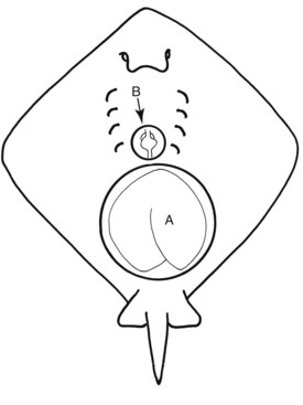
Rays have gill slits on their ventral body wall and usually a spiracle (respiratory aperture) on their dorsum. Respiration occurs through active pumping of water over gills by pulling water into the mouth or the spiracles and then bathing the gills; thus, for those species that rest on the bottom, use of the spiracles provides unobstructed respiration, unlike some of their shark counterparts that must continually swim to respirate.8 Manta rays are unique in that they possess gill rakers, much like the whale shark. The musculoskeletal system is almost completely cartilaginous, with the exception of occasional ossification of the vertebral column. The simple cardiovascular system has one ventricle, one atrium, a sinus venosum, and a conus arteriosis (Fig. 22-1). The heart is separated from the coelom by a rigid pericardium.16 A renal portal system has been documented. Rays possess no bone marrow, so hematopoiesis occurs via the spleen, Leydig organ, and epigonal organ. The latter two vary greatly in size and contribution of hematopoietic cells per species. Thyroid tissue is present but is disseminated and difficult to identify unless diseased.
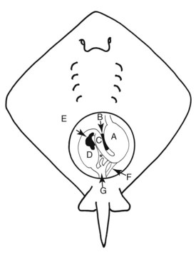
The liver is a predominant organ, occupying most of the length and width of the coelom (see Fig. 22-1). It is light tan in color, a reflection of the high lipid content (a natural hepatic lipidosis); lipids make up to 80% of the liver’s architecture.8 The liver contributes significantly to buoyancy because there is no swim bladder. A gallbladder is present that produces bile acids that form alcohol sulfate esters (versus taurine salts in birds and mammals, which are reflected in available bile acid assays). The gastrointestinal tract (Fig. 22-2) is simple, with a distensible esophagus, descending cardiac stomach with pronounced rugae, and ascending pyloric stomach that is connected directly to a spiral colon, also called the spiral valve. The colon varies anatomically with species but typically has copious folds or layers that increase digestive capability. There is a rectum that empties into a cloaca. Unique to elasmobranchs is the rectal gland, which opens into the intestine-rectum area and is involved in the osmoregulation of sodium and chloride ions. Freshwater stingrays have reduced or no functional rectal gland tissue, depending on their water salinity.2 Coelomic (abdominal) pores, adjacent to cloaca, have a direct connection to the coelomic cavity. Their function is not fully understood but is suspected to regulate electrolytes and maintain osmoregulatory homeostasis.6 Some clinicians have used these pores for examination of the coelom with a laparoscope or for flushing the coelom; others question this technique because of its suspected regulatory function.
Vision is poor compared to other senses; pupil dilation may be controlled by the animal, unlike in teleosts but compared with mammals’ pupillary light reflexes, are slow or difficult to assess when stimulated. Because of pigmentation, the lenses in some species may be slightly opaque.4 Olfaction and taste are keen sensory abilities. Non–filter-feeding rays mouth objects to determine whether they are edible and they may discriminate between fish types. It may be difficult to distinguish mouthing or crushing of food from actual consumption of food. The ampullae of Lorenzini (mucopolysaccharide-filled electrosensory organs) detect external vibration and low-frequency electrical pulses for the detection of animate versus inanimate objects.8 Filters and pumps too close to an enclosure may disturb these organs and cause aberrant behavior. A lateral line is present, with a lining of neuromast cells that respond to pressure changes.
All saltwater rays are hyperosmotic to their environment. They have a high urea content in the blood as well as other solutes that increase osmolarity. A specialized protein, trimethylamine N-oxide (TMAO), is lighter than seawater and is thought to aid in buoyancy.8 Freshwater elasmobranchs have a significantly lower urea content; those that are obligate freshwater animals have negligible levels.2
Copulation in the rays occurs with the male grasping the female on the wings (usually resulting is some degree of trauma) and then inserting a clasper into the female’s cloaca, which transfers sperm from the male. Sperm storage occurs in multiple species with varying durations. Aplacental viviparity occurs with the production of uterine milk, which is absorbed by the embryo. The uterus develops trophonemata; these are villi of the uterine epithelium that secrete histotroph, a rich yellow opaque fluid that feeds the embryos (but should not be mistaken for pyometra). Ray embryos have a yolk sac, but it is absorbed by the time gestation is 75% complete, after which they rely on histotroph for nutrition. Gestation is variable (2 to 12 months) and estimating the birth date may be complicated, given their ability to store sperm for several months.4,13
Husbandry and Management
Water quality parameters and specific exhibit suggestions may be found elsewhere.13 Special focus will be paid to contact or touch pools because these have gained immense popularity in the last several years. There has been some notoriety associated with these exhibits in the media that has reported on large population declines caused by sudden animal death. In many touch pools, these are related to a life support problem. Most of these pools are shallow and populated beyond optimum carrying capacity. Many of these exhibits are temporary and housed in facilities that do not typically support large aquaria. Specific causes of these events have been attributed to hyperthermia, hypoxia, well water contamination by heavy metals, and inanition caused by poor acclimation. The first two issues may be avoided with alarm systems that broadcast an alert when water quality parameters are out of range. Water sources should be tested for contaminants. Acclimation becomes an issue with recent capture of free-range specimens, usually young animals that need special attention to transition to a captive diet. Using experienced adult animals in these exhibits may avoid this issue; alternately, the animals must be managed throughout this period by offering foods that approximate their wild diet and often by supportive assist feeding. Rarely do these animals suffer from touch-related issues. Although these animals are encouraged to be out and touchable, hiding places should be made available for them. Most will choose to interact because the species chosen are gregarious by nature.
Restraint
Not all rays possess spines or barbs on their tails; for example, Urogymus spp. and the manta rays lack barbs and are not venomous.5 Those that do have spines or barbs are typically gentle but will defend themselves in situations in which they are stepped on or manipulated excessively. Although being abraded or punctured by a stingray barb is dangerous, one should also be cautious of their bites, recalling that these animals crush crustacean prey. The barb or spine of a stingray is cartilaginous and often serrated. Venom on the barbs is not produced by venom glands but by two longitudinal grooves under the barb that contain venom-secreting glandular cells. The spine is covered with epidermis; when a human is punctured, not only is there venom to contend with but pieces of integument and cells remain in the wound, increasing the exposure to microorganisms and venom. The venoms are largely cardiotoxic, although some are neurotoxic as well.
Protocols for managing stingray injuries are recommended.5 Some institutions will trim the barbs of these animals, which reduces the risk of puncture, but exposure through abrasion is still possible because the base of the barb still contains venom. Barbs are naturally shed two or three times/year; therefore, regrowth may be expected at this interval.13 Removal of the barb may be done surgically; on occasion, the procedure may result in infection, permanent scarring, or regrowth, sometimes in an abnormal position. In freshwater stingrays, the epidermal bumps around the barb also contain venom. Anecdotally, freshwater stingrays have more potent venom, resulting in a greater pain response in affected humans, although most of these injuries are manageable with analgesics. Placing plastic tubing around the tail and barb will prevent accidental exposure.
Manual restraint is possible, although special precautions should be used, as noted. Tonic-clonic immobilization may be achieved by flipping the animal over until it is in a trancelike state; care should be taken when placing pressure on the coelom, because the liver may fracture. This is much easier with animals that are acclimated to handling; for them, operant condition is a successful tool for rays. Anesthesia should be used in naïve or large specimens and for lengthy procedures. Chemical restraint is usually accomplished by immersion. Injectables are not commonly used and no reports have been published. Most anesthesia is performed via bathing in tricaine methane sulfonate (MS-222), with an equal mass of buffer (bicarbonate), in a variety of concentration ranges, from 60 to 300 ppm (typically at the lower end of that dose range). Animals should be ventilated with a syringe and tubing or a pump delivering water that is slowly and gently flowed over the gills; a rapid current may prevent gas exchange. Recoveries should also be under gentle flow ventilation because this greatly increases the oxygen saturation of the blood rather than the traditional walking of the animal in the water. In a recent study of different immersion anesthetics, 20 ppm eugenol, 15.7 ppm isoeugenol, and 55 to 65 ppm MS-222 were compared in Daysatis americana. Minor variations were noted but, overall, all the agents provided adequate anesthesia for short-term immobilizations and minor procedures.11 An important note when handling gravid females is that abortion may occur with anesthesia or handling, more likely later in gestation. If very late in gestation, it is possible that these pups may be successfully raised.
Stay updated, free articles. Join our Telegram channel

Full access? Get Clinical Tree


