Oliver D. Pynn
Managing Dystocia in the Field
Dystocia or difficult parturition represents a true emergency faced by the equine veterinarian, and effective management is essential for survival of both foal and mare and for the mare’s subsequent fertility. Dystocia is most commonly caused by abnormalities of fetal presentation (the portion of the fetus that enters the vaginal canal first), position (the relationship of the fetal spine to the mare’s pelvis), or posture (the relationship of the fetal extremities to its body), but can also result from fetal oversize or maternal factors such as pelvic abnormalities or uterine inertia. The incidence of dystocia has been reported to be anywhere from 4% to 10% of all births. Dystocia should be approached in a systematic way, with the aim of delivering a live foal as quickly as possible with minimal complications for the mare.
An understanding of the normal time course and progression of foaling is essential. Most important, the second stage of labor, which starts with rupture of the chorioallantoic sac and ends with delivery of the foal, should take 20 minutes or less. Two key indications that a mare is having difficulty are failure of the amniotic membrane or any fetal body parts to appear at the vulval lips after 5 minutes from the onset of second-stage labor and failure of delivery to progress. Obviously, any abnormality of fetal parts showing at the vulval lips also warrants immediate investigation. Failure to quickly identify dystocia can lead to impaction of the fetus against the pelvis and make correction more difficult. Delay in second-stage labor of more than 30 to 40 minutes is associated with a significant increase in foal morbidity and death. When the chorioallantois does not rupture, resulting in a “red bag” presentation, there will have been sufficient placental separation to cause fetal hypoxia, so foaling attendants should be instructed to rupture the membranes and help deliver the foal.
Diagnosis
A thorough physical examination of a mare with dystocia is often not practical, especially if the mare is recumbent. However, a rapid assessment for signs of shock or hemorrhage is important. It is also important to get a brief history, including the expected foaling date and a timeline for the dystocia. Care should be taken with foaling mares, because they can be fractious and unpredictable. Examination in a large foaling box is preferable, and additional restraint can be achieved with a nose twitch. The mare can also be sedated with a combination of xylazine (0.5 mg/kg, IV) and butorphanol (0.02 mg/kg, IV). The sedative will transfer to the fetus and also may reduce placental blood flow, further compromising a hypoxic fetus, so xylazine is preferable to any of the other α2 agonists because of its shorter action. Epidural anesthesia can be used to achieve perineal analgesia and reduce straining, but it is time consuming to perform and carries a risk for hindlimb weakness and ataxia, which may be problematic if the horse is to be transported to a surgical facility. However, it can be useful in the field to aid manipulation, especially if the foal is dead. For epidural administration, a combination of xylazine (0.17 mg/kg) and mepivacaine (0.15 mg/kg) can be used, made up to 10 mL with sterile saline solution (e.g., 1 mL of 10% xylazine, 4 mL of 2% mepivacaine, and 5 mL of sterile saline). The volume used is adjusted according to the body weight of the horse, but the full 10-mL volume would be used for a 550-kg mare.
Before vulval or vaginal examination, the tail should be wrapped and the perineal region thoroughly cleansed, because the importance of a high level of hygiene cannot be overemphasized even though time is of the essence in this situation. The obstetrician’s hands and arms should also be cleaned, and it is preferable to wear long rectal sleeves to minimize abrasive damage to the uterus, cervix, and vagina. Donning a pair of surgical gloves over rectal sleeves provides better sensation and grip. To aid examination by inducing uterine relaxation, clenbuterol (0.8 µg/kg, IV) can be given. Copious lubrication is also essential, and a sterile stomach tube and pump can be used to instill a mixture of water and lubrication into the uterus. It is important to determine the posture, position, and presentation of the fetus, fetal size relative to the mare’s pelvic canal, and fetal viability. Fetal oversize is not a common issue in horses because the size of the mare’s uterus has more effect on foal size than does stallion size. However, relative fetal oversize can still sometimes be a factor, particularly in maiden mares. Determining whether a fetus is dead or alive often has a bearing on the course of action taken. This can be difficult because fetal reflexes are dulled, especially if the fetus is impacted against the pelvis. Detection of the fetal heartbeat by palpating its thorax may not be possible, and in such cases, transabdominal ultrasonography of the fetal heart with a 3.5-MHz probe can be used.
Dystocia is generally resolved in one of the following ways: by assisted vaginal delivery in a standing or recumbent mare; controlled vaginal delivery (CVD), whereby the mare is anesthetized briefly to aid mutation of the fetus; fetotomy, whereby a dead fetus can be delivered in multiple parts; and cesarean section, wherein the fetus is removed from the uterus surgically. Time is of particular importance, and a quick decision regarding how to proceed is essential. Economic constraints, a bias of value placed on the mare or on the foal, the expertise of the clinician, and the proximity of a good surgical center and neonatal intensive care unit should all be considered. Assisted vaginal delivery is the preferred option in any case of dystocia because there is no need for general anesthesia or surgical intervention, which both carry inherent risks to mare and foal. However, vaginal manipulations can be very difficult because of inadequate space in the pelvic canal and long fetal extremities, and excessive intervention carries the risk for trauma to the mare’s reproductive tract and impaired future reproductive health.
Abnormalities of Fetal Position
Malposition with the fetus upside-down (i.e., in dorsopubic position) is a common presenting complaint and usually represents a delay in the foal’s turning into the normal position for foaling. This can often be self-corrected by allowing the mare to stand up and lie down again, but applying pressure over the fetus’ shoulder can encourage correction if assistance is required. Although it is rare, the possibility of uterine torsion should be considered if the fetus is in this position. Diagnosis is made by palpation of the broad ligaments per rectum. Most of these cases can be corrected by grasping the fetus ventrolaterally through the cervix and rocking it until the fetus and uterus roll into the correct position.
Abnormalities of Fetal Posture
Foot-nape posture occurs when the fetal forelimb or forelimbs lie over its head, which if left uncorrected can lead to injury to the dorsal aspect of the vagina and even to creation of a rectovaginal fistula or third-degree laceration. It is relatively easily corrected by repulsion of the fetus, which enables repositioning of the limb or limbs laterally into the normal position.
Elbow lock occurs when the fetal elbows become impacted against the pelvic brim. The fetus should be repelled and dorsal and medial traction applied to the affected limb, which will release the elbow and allow full extension of the limb.
Carpal flexion can occur unilaterally or bilaterally and is the likely reason for one or both limbs not being visible at the vulval lips. The fetus should be repelled to allow for correction. A foaling rope placed around the pastern is useful and enables control of the limb. The flexed knee can then be repelled and deviated laterally, which allows the foot to be manipulated medially and brought into the correct position. The obstetrician’s hand should cup the foot of the fetus during manipulations so that the uterus is protected from injury. If correction is proving difficult, fetal carpal contracture should be considered. This is the most common congenital defect of the equine fetus and usually occurs bilaterally. Attempts to deliver these foals per vagina, without fetotomy, risk significant injury to the reproductive tract.
Head and neck deviations occur either laterally or ventrally. Deviations can occur as a result of a viable fetus retracting its head away from vaginal manipulations. To prevent this, a foaling rope can be placed over the fetus’ head and through its mouth prior to initiation of manipulations. With ventral deviation, the head usually deviates just below the pelvic brim, but the depth to which it deviates can vary. If the lower jaw can be snared, the head can be repelled and then brought up. Care must be taken to avoid fracture of the jaw. If ventral deviation of the head and neck occurs with bilateral carpal flexion, the head and neck flexion should be corrected before correction of the limbs. Lateral deviation of the head and neck can be much more difficult to correct, and in the author’s experience, CVD is much more rewarding than prolonged manipulation without general anesthesia. Wry-neck deformity should be considered in cases refractory to corrective attempts.
Dog-sitting or a ventrovertical presentation occurs when one or, less commonly, both hindlimbs become caught on the brim of the pelvis underneath the fetus and results from unilateral or bilateral hip flexion. This usually manifests as an apparently normal foaling that has failed to progress beyond the level of the fetus’ chest. Fetal hypoxia and death usually occur rapidly because of compression of the umbilical cord. In these cases, sedation of the mare is usually essential because mares are often extremely fractious. If the foal is still alive, the ex utero intrapartum treatment (EXIT) procedure (see later) should be considered before attempting manipulations. With the aid of copious volumes of lubrication pumped around and behind the foal, the offending hind foot can sometimes be palpated at the pelvic brim. This can sometimes be repelled and, with an assistant applying traction to the fetus, may fall back into the normal position and enable vaginal delivery. Often CVD is required, especially if excessive traction has been applied to the fetus and it has become impacted in the pelvic canal. Injury of the ventral aspect of the mare’s cervix is a common sequel to this presentation.
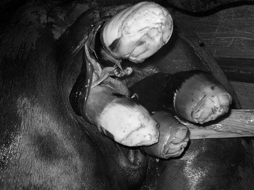
The incidence of posterior presentation is very low. However, when it does occur, fetal morbidity or death is likely secondary to hypoxia as a result of early compression or severance of the umbilical cord. If the hind limbs are presented, gentle traction usually allows for normal vaginal delivery. The risk for compression of the umbilical cord is high, and delivery must proceed quickly to prevent fetal hypoxia. Hock flexion and hip flexion are very difficult to correct, even with CVD, and a cesarean section provides the best outcome for mare and foal. The same can be said for transverse presentations, which occur even less frequently. With a ventrotransverse presentation (Figure 170-1), four feet will be presented at the vulval lips, a situation that must be differentiated from the rare event of a twin foaling.
< div class='tao-gold-member'>
Stay updated, free articles. Join our Telegram channel

Full access? Get Clinical Tree


