Magnetic Resonance Imaging

Chapter 21 Magnetic Resonance Imaging


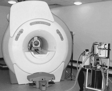
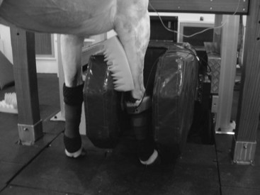
Magnetic resonance imaging (MRI) is a method of imaging that can allow examination of both osseous and soft tissues with detailed anatomical resolution and also provides some physiological information. Advances in technology during the past 10 years have made it possible for MRI to become a practical diagnostic tool in both anesthetized and standing horses (Figures 21-1 and 21-2).1 Use of MRI has revolutionized our understanding of foot pain in the horse and has considerably improved understanding of other types of lameness further proximally in the limb.2-5 As a result, MRI is increasingly being used for diagnosis of orthopedic disorders in equine practice and can add considerable information to a diagnostic investigation. However, as with any imaging technique, MRI is only useful in the context of the entire clinical picture and needs to be interpreted in light of other diagnostic findings. Terminology is sometimes confused: magnetic resonance (MR) refers to the absorption of energy exhibited by particles (as atomic nuclei or electrons) in a static magnetic field when the particles are exposed to electromagnetic radiation of certain frequencies. MRI refers to a noninvasive diagnostic technique that produces computerized images of internal body tissues and is based on nuclear magnetic resonance of atoms within the body induced by the application of radio waves.


Production of Magnetic Resonance Images


MR images are produced through interactions between a strong magnet, a radiofrequency (RF) coil, magnetic field gradients, and powerful computing facilities. MR images are based on the spinning motion of MR active nuclei (nuclei with an odd number of protons). Hydrogen nuclei are used in clinical MRI because they are abundant in the body and are particularly mobile in fat and water. When a horse (or body part) is put into the magnet, the MR active nuclei align parallel or antiparallel to the static magnetic field. To produce an image, an RF wave at a specific frequency (dependent on the magnet field strength and the MR active nuclei selected) is applied at 90 degrees to the static magnetic field, B0. This gives energy to the hydrogen nuclei, which then spin faster and alter the net magnetization vector away from the static magnetic field. When the RF wave terminates, energy is lost from the excited hydrogen nuclei, either to the surrounding environment (spin-lattice or T1 relaxation) or to adjacent nuclei (spin-spin or T2 relaxation), and the net magnetization vector returns to zero. This energy loss is detected by the receiver as MR signal. The signal is then converted to a digital image using complex computer software. Each pixel within the matrix has a particular signal intensity, and the accumulated pixels make up the final image.


Magnets may be considered high-, mid-, and low-field strength, with field strength measured in Tesla. Magnetic field homogeneity is important in production of good quality images. RF coils are made to surround the region of interest with as little space between the coil and the area of interest as possible to maximize signal detection. They may be both a transmitter (transmits the RF waves) and receiver (detects the MR signal produced). The magnetic field gradients are used to place the MR signal in space. This is achieved by generating short-term spatial variation in magnetic field strength across the horse (or body part). There are three sets of gradient coils, each oriented in a different direction. Repeated application of large electrical current pulses results in the gradient coils making a loud noise during scan acquisition, especially at higher field strengths.


A pulse sequence includes a defined series of RF waves, gradient maneuvers, and signal detection. The repetition time (TR) defines the time between repetitions, and the echo time (TE) defines the time between the RF pulse and detection of the echo (MR signal). Selection of pulse sequence determines the image contrast and appearance of particular tissues.


Images are produced in a gray scale with the contrast defined by the particular pulse sequence used for image acquisition, the imaging parameters, and the properties of the tissue being imaged. Information is acquired as tomographic slices, so the orientation and thickness of slices (or three-dimensional [3D] volume datasets) can be varied and defined.


Generally a pilot scan with wide slice thickness is performed in three planes, and further image sequences are oriented and positioned relative to the pilot scan. It is normal practice to acquire images in three planes: sagittal, dorsal (coronal), and transverse (axial).


A complete MRI scan of a horse usually includes a series of different pulse sequences, the choice of which is determined by the operator. The tissues being examined, the suspected pathology, and the type of MRI system influence the selection of pulse sequences. For horses, the choice of sequence is also influenced greatly by time constraints to minimize risk of movement in a standing horse and limit time under general anesthesia.


Various manipulations during image acquisition can be used to look at particular features of tissues, such as fat suppression techniques to identify pathology in medullary bone. Intravenous administration of gadolinium contrast medium can be used to demonstrate blood flow, perfusion of a suspect area, or to investigate damage to the blood–brain barrier. Postprocessing manipulations can be used to reorient images acquired in an oblique orientation; to measure size, area, or volume; for motion correction; or to further understand signal intensity patterns.


Equipment for MRI in Horses


MRI can be performed in horses using various different types of systems. High-field closed systems are similar to those used in many human hospitals. These units have a superconducting, helium-cooled magnet in a cylindrical configuration into which the area of interest of the horse has to be squeezed; thus the horse needs to be under general anesthesia, and a team of operators is required to position the horse (see Figure 21-1). There are low-field open MRI systems that also require horses to be imaged under general anesthesia but have fewer restrictions on space; therefore it may be easier to position the horse into the imaging portion of the magnet. A low-field MR system for the standing horse has been developed specifically for the equine market, which has allowed images to be acquired with horses under sedation (see Figure 21-2). Use of motion-insensitive imaging sequences helps to account for slight sway that may occur during sedation. Time for image acquisition is generally longer in low-field units for the same or less resolution and image quality as a high-field system; thus horses may require sedation or general anesthesia for more prolonged periods to acquire images of the entire region required.


For the MRI systems available, in general the areas that can be imaged in the adult horse are limited to the head and cranial aspect of the neck, the forelimb up to and including the carpus, and the hindlimb up to and including the tarsus, although images of the stifle can also be obtained in a few systems in horses of suitable size and conformation.6 In a foal, it may be possible to place the entire body into the magnet in a high-field closed or low-field open system.


Jun 4, 2016 | Posted by in EQUINE MEDICINE | Comments Off on Magnetic Resonance Imaging

Full access? Get Clinical Tree

Get Clinical Tree app for offline access