M
Macadamia Nut Toxicosis
BASIC INFORMATION 
DEFINITION
Acute and often self-limiting toxicosis of dogs resulting from ingestion of macadamia nuts. It is characterized by paresis, depression, vomiting, ataxia, tremors, hyperthermia, abdominal pain, lameness, and/or stiffness. Toxicosis can occur after ingestion of commercially available macadamia nuts or macadamia nut-containing cookies or candies.
CLINICAL PRESENTATION
DIAGNOSIS 
DIAGNOSTIC OVERVIEW
Diagnosis is based entirely on history and physical signs: observed or suspected ingestion of macadamia nuts, especially if concurrent signs (paresis, ataxia, stiffness, hyperthermia, and/or vomiting within 12 hours of exposure) are present, is sufficient to warrant treatment.
DIFFERENTIAL DIAGNOSIS
TREATMENT 
TREATMENT OVERVIEW
Treatment is general and supportive: induction of emesis and administration of activated charcoal if ingestion is suspected or confirmed and patient is stable. Management of complications of severe intoxications (e.g., nursing care if recumbent; treatment of hyperthermia) is applied as needed. Most clinical signs resolve within 24-48 hours with or without treatment.
ACUTE GENERAL TREATMENT
PEARLS & CONSIDERATIONS 
COMMENTS
Malassezia Dermatitis
BASIC INFORMATION 
DEFINITION
An extremely common pruritic dermatosis caused by the overgrowth of Malassezia spp. yeast. Most infections are caused by the lipophilic unicellular organism Malassezia pachydermatis.
EPIDEMIOLOGY
SPECIES, AGE, SEX: Very common in dogs of all ages; uncommon in cats of any age; either sex
GENETICS & BREED PREDISPOSITION: May occur in any breed of dog or cat. Terriers, shih tzus, dachshunds, shar-peis, spaniels, hounds, and German shepherd dogs seem predisposed, likely to underlying conditions favorable to yeast overgrowth. Devon Rex cats are more susceptible.
RISK FACTORS
CONTAGION & ZOONOSIS: Malassezia yeasts have been transmitted from the contaminated hands of dog-owning health care workers to infants in an intensive care nursery, causing mycotic sepsis. Therefore Malassezia yeast should be considered a potential zoonotic agent, especially in immunoincompetent individuals.
GEOGRAPHY AND SEASONALITY: Malassezia dermatitis may occur more frequently in humid geographic regions, and/or with underlying causes that worsen seasonally (e.g., atopic dermatitis).
CLINICAL PRESENTATION
DISEASE FORMS/SUBTYPES
HISTORY, CHIEF COMPLAINT: Intense pruritus: most common chief complaint. Rancid offensive odor; an oily coat; hair loss (alopecia); redness (erythroderma); thickened, elephant-like skin (lichenification); scaling (dander); and/or relapsing and remitting dermatitis unresponsive to antibiotics or glucocorticoids are also frequently noted.
PHYSICAL EXAM FINDINGS

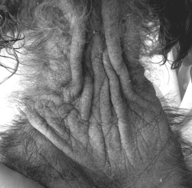
(Copyright Dr. Manon Paradis.)
ETIOLOGY AND PATHOPHYSIOLOGY
Malassezia spp. yeasts are part of the normal skin microflora. They become opportunistic invaders when changes occur in the cutaneous microclimate (e.g., lipid composition, relative humidity) or defense mechanisms (e.g., epidermal barrier dysfunction, immunosuppression). Once colonization takes place, yeasts may release proteases and lipases that alter cutaneous homeostasis, allowing for continued yeast overgrowth. In some atopic dogs with cytologic demonstration of yeasts, Malassezia may elicit a type-1 cutaneous hypersensitivity reaction.
DIAGNOSIS 
DIAGNOSTIC OVERVIEW
Malassezia dermatitis should be considered in any pruritic dog or cat. Confirmation requires typical clinical signs, cytological demonstration of yeast, and most importantly, response to antifungal therapy. Underlying primary conditions must be identified and corrected. Importantly, Devon Rex cats may not have an identifiable predisposing disease.
DIFFERENTIAL DIAGNOSIS
Since Malassezia dermatitis is a secondary complication of underlying disorders in most cases, the chief differential diagnosis consists of identifying whether the underlying disorder (see Risk Factors, above) exists in an uncomplicated state or if secondary Malassezia dermatitis is present.
INITIAL DATABASE
ADVANCED OR CONFIRMATORY TESTING
TREATMENT 
TREATMENT OVERVIEW
The main goal your job is to reduce pruritus and remove seborrhea. This is accomplished by killing yeast and controlling for underlying primary diseases and risk factors.
ACUTE GENERAL TREATMENT
CHRONIC TREATMENT
DRUG INTERACTIONS
Azole therapy may alter the metabolism or distribution of other prescribed medication by inhibiting cytochrome P450 metabolizing enzymes and P-glycoprotein transporting pumps. Terbinafine does not inhibit these enzymes. Specifically, azoles cannot be given with macrocyclic lactones (e.g., avermectins); if so, dosage reduction and close monitoring is required (see p. 706).
POSSIBLE COMPLICATIONS
PROGNOSIS AND OUTCOME 
PEARLS & CONSIDERATIONS 
COMMENTS
PREVENTION
Correctly identifying and treating for all underlying predisposing factors favorable to yeast overgrowth
Åhman S, Perrins N, Bond R. Treatment of Malassezia pachydermatts-associated seborrheic dermatitis in Devon Rex cats with itraconazole-a pilot study. Vet Dermatol. 2007;18:171-174.
Chen TA, Hill PB. The biology of Malassezia organisms and their ability to induce immune responses and skin disease. Vet Dermatol. 2005;16:4-26.
Negre A, Bensignor E, Guillot J. Evidence-based veterinary dermatology: a systematic review of interventions for Malassezia dermatitis in dogs. Vet Dermatol. 2008;20:1-12.
Malignant Fibrous Histiocytoma
BASIC INFORMATION
DEFINITION
An uncommon primary tumor made up of fibrous and inflammatory cells thought to arise from a primitive mesenchymal cell. It is not to be confused with malignant histiocytosis (histiocytic sarcoma), a disease of localized or multisystemic histiocytic infiltration (see p. 535).
EPIDEMIOLOGY
SPECIES, AGE, SEX: Malignant fibrous histiocytoma is an uncommon tumor of the skin and subcutaneous tissue of dogs and cats, and can also occur in the spleen in dogs. This tumor type has been reported at injection sites in cats (see p. 610).
GENETICS & BREED PREDISPOSITION: Golden retrievers and rottweilers may be overrepresented.
CLINICAL PRESENTATION
DISEASE FORMS/SUBTYPES
HISTORY, CHIEF COMPLAINT
ETIOLOGY AND PATHOPHYSIOLOGY
DIAGNOSIS
DIAGNOSTIC OVERVIEW
Definitive diagnosis can only be confirmed via histopathologic analysis, although additional testing such as diagnostic imaging is often helpful in defining the extent of the tumor.
INITIAL DATABASE
ADVANCED OR CONFIRMATORY TESTING
TREATMENT
TREATMENT OVERVIEW
Definitive treatment is based on complete eradication of the primary tumor whenever possible. However, additional treatment such as chemotherapy may be indicated to prevent or delay metastases or in dogs with high-grade tumors, the giant-cell variant of this tumor or tumors that have already metastasized. Palliative treatment options, such as palliative radiation, may help control pain or discomfort in patients with advanced tumors or in patients where definitive treatment cannot be tolerated.
ACUTE AND CHRONIC TREATMENT
POSSIBLE COMPLICATIONS
Complications of treatment depend on types of treatments and location of the primary tumor.
RECOMMENDED MONITORING
After appropriate local treatment, follow-up examination should be done routinely to monitor for recurrence (every 2-3 months) and metastasis (thoracic radiographs at 6 months and 1 year). High-grade tumors may require more frequent monitoring (at least every 2-3 months) for metastases during and after chemotherapy administration.
PROGNOSIS AND OUTCOME 
PEARLS & CONSIDERATIONS 
COMMENTS
Aside from the giant-cell variant, malignant fibrous histiocytoma should be considered like other soft tissue sarcomas (see p. 1034) in treatment and prognosis. Tumors at injection sites in cats should be treated like injection-site sarcomas (see p. 610).
Malnutrition
BASIC INFORMATION 
DEFINITION
Malnutrition is the inappropriate intake of nutrients, resulting in nutritional deficiencies or excesses.
EPIDEMIOLOGY
SPECIES, AGE, SEX: Malnutrition can affect animals of any species, age, life stage, or lifestyle.
GENETICS & BREED PREDISPOSITION: No specific genetic link or breed disposition is known for malnutrition associated with nutritional deficiencies. An increased prevalence of obesity resulting from excessive caloric intake has been associated with basset hounds, beagles, Cairn terriers, Cavalier King Charles spaniels, cocker spaniels, long-haired dachshunds, Labrador retrievers, and Shetland sheepdogs.
RISK FACTORS
GEOGRAPHY AND SEASONALITY: Inadequate food or water intake during severe weather conditions may result in nutrient deprivation.
CLINICAL PRESENTATION
HISTORY, CHIEF COMPLAINT
PHYSICAL EXAM FINDINGS: Nutrient-deprived animals are often in poor physical appearance: thin body condition; dry, coarse haircoat; flaky skin; hyperkeratosis; muscle wasting; broken or missing teeth; skeletal abnormalities; pressure sores; poor wound healing. Obese animals are also in poor physical condition with fat deposition over the ribs, hips, tail head, and throughout the abdomen. Some are unable to adequately groom themselves, which can result in an unkempt hair coat, dry skin, skinfold dermatitis, and the inability to walk, run, or jump.
ETIOLOGY AND PATHOPHYSIOLOGY
DIAGNOSIS 
DIAGNOSTIC OVERVIEW
The diagnosis of malnutrition is made on the basis of a thorough diet history and the physical examination findings.
DIFFERENTIAL DIAGNOSIS
Gastrointestinal disease: protein-losing enteropathy, lymphocytic-plasmacytic enteritis, inflammatory bowel disease
INITIAL DATABASE
TREATMENT 
TREATMENT OVERVIEW
The goals of treatment are to correct nutrient deficiencies or excesses and return patient to a normal plane of nutrition and a more appropriate body weight and body condition.
ACUTE GENERAL TREATMENT
CHRONIC TREATMENT
DRUG INTERACTIONS
Medical therapies instituted for primary conditions will be altered without adequate nutrition support.
PROGNOSIS AND OUTCOME 
PEARLS & CONSIDERATIONS 
COMMENTS
PREVENTION
TECHNICIAN TIPS
Abood S, McLoughlin M, Buffington T. Enteral nutrition. In: DiBartola SP, editor. Fluid, electrolyte, and acid–base disorders small animal practice. ed 3. St Louis: Saunders Elsevier; 2006:601-620.
Buffington T, Holloway C, Abood S. Manual of veterinary dietetics. St Louis: Elsevier, 2004.
Chan DL. Nutritional requirements of the critically ill patient. Clin Tech Small Animal Pract. 2004;19:1-5.
Eirmann L, Michel K. Enteral Nutrition. In: Silverstein D, Hopper K, editors. Small animal critical care medicine. St Louis: Saunders, 2009.
Freeman L, Chan D. Total parenteral nutrition. In: DiBartola SP, editor. Fluid, electrolyte, and acid–base disorders small animal practice. ed 3. St Louis: Saunders Elsevier; 2006:548-600.
Remillard RL, Armstrong PJ, Davenport DJ. Assisted feeding in hospitalized patients: enteral and parenteral nutrition. In Hand MS, Thatcher CD, Remillard RL, Roudebush PR, editors: Small animal clinical nutrition, ed 4, Topeka: Mark Morris Institute, 2000.
Malnutrition, Home-Prepared Diets
DEFINITION
Feeding of an unbalanced diet of uncooked and cooked foods as a sole source of nutrition; this practice is becoming more common among pet owners.
EPIDEMIOLOGY
SPECIES, AGE, SEX: Dogs more likely to be fed home-prepared diets than cats. Young and reproductive animals at increased risk for deficiencies and excess. All animals at risk of illness from foodborne pathogens.
GENETICS & BREED PREDISPOSITION: Dogs and cats: all breeds affected. Dogs: large and giant breeds at increased risk of developmental orthopedic disease. Cats: taurine-deficient cardiomyopathy and/or central retinal degeneration. CONTAGION & ZOONOSIS: Dogs and cats: Salmonella, Escherichia coli, Campylobacter, Toxoplasma, Cryptosporidium
ASSOCIATED CONDITIONS & DISORDERS: Poor skin and coat, obesity, vomiting, diarrhea, pathologic fractures, seizures, pancreatitis (high-fat diet), oral trauma, intestinal obstruction/perforation

Full access? Get Clinical Tree

