M
Magnetic Resonance Imaging Scan
OVERVIEW AND GOALS
• Magnetic resonance imaging (MRI) is a method of cross-sectional imaging that does not involve ionizing radiation. When compared to radiography, MRI allows improved evaluation of areas of complex anatomy by avoiding superimposition of multiple structures. The contrast resolution of MRI is vastly superior to that of radiography and computed tomography (CT).
CONTRAINDICATIONS
• Presence of a pacemaker: in a minority of patients, the MRI magnetic field may alter the pacemaker program or cause the pacemaker to oscillate in the patient. In human medicine, MRI suites bear warnings forbidding entry to any person with a pacemaker, so it is unlikely that an MRI technician will allow a veterinary patient with a pacemaker to be scanned.
GENERAL MRI SAFETY CONSIDERATIONS:
• Injury to the patient and/or damage to the MRI magnet can occur if careful attention is not paid to basic MR safety as regards the presence of metallic objects within the MRI suite.
• The MR magnet is the core of the MRI unit, and the magnet is always “on,” 24 hours a day. Therefore, it is never safe to have loose metallic objects in the vicinity of the magnet—that is, anywhere in the same room as the MRI unit (MRI suite).
• Since the chemical composition of metal objects (ferrous or nonferrous) may not be known, all metallic objects should be regarded as potential safety hazards and excluded from the MRI suite.
• All equipment used in the area of the MRI magnet must be constructed of plastic and/or nonferrous metal or must be kept a safe distance from the magnet and secured. Recorded instances where this guideline was not followed have produced injury and death in patients and hospital personnel through blunt trauma caused by large objects which transform into projectiles owing to their strong attraction to the magnet.
• The safe distance from the magnet will depend on the field strength of the magnet, the shielding of the magnet and the amount of ferrous metal in the object. A distance of 36 inches (3 meters) is generally sufficient for a magnet of 1.0 tesla or less field strength.
EQUIPMENT, ANESTHESIA
• Emergency kit:
○ As with any procedure performed under general anesthesia, the equipment and drugs necessary for emergency cardiopulmonary cerebral resuscitation (CPCR) should be immediately available.
• Paramagnetic agents:
○ Gadodiamide: Omniscan
▪ Nephrogenic systemic fibrosis has been reported as a sequela to the injection of intravenous paramagnetic contrast agents in human patients with renal insufficiency. This syndrome has not been reported in veterinary patients, but care should be taken when administering these agents to patients with renal dysfunction. Gadodiamide has a higher safety ratio than Gd-DTPA.
ANTICIPATED TIME
The time required to perform the study depends on many factors:
• Field strength of the magnet: the higher the field strength of the magnet, the more quickly the study can be performed.
• Number and type of imaging sequences performed: unlike CT, MRI involves multiple separate image acquisitions. Images are obtained in multiple planes and using multiple different imaging parameters; the more acquisitions performed, the longer the study.
APPROXIMATE TIMES (Based on use of a 1.0-tesla magnet):
PREPARATION: IMPORTANT CHECKPOINTS
• Obtain thorough patient history: pacemaker? nonferrous metallic implants/foreign bodies? ferrous metallic implants/foreign bodies?
• Many patients undergoing MR imaging are older and may have multiple diseases. Routine laboratory evaluation and thoracic and abdominal screening may be indicated in these patients for two reasons: to rule out the presence of concurrent disease that may affect the patient’s ability to tolerate general anesthesia and to identify the presence of any potentially life-shortening disease process unrelated to the primary complaint (e.g., “incidental” splenic mass).
• Make sure all needed equipment is available and functional. This is especially important if the study is to be performed at an outside (e.g. nonveterinary) facility.
• Preparation for general anesthesia:
○ Place IV catheter: when placing the catheter, the anticipated position of the patient within the MRI unit must be considered, and the catheter should be placed to allow easy access to it during the procedure. For example, in patients undergoing MR scan of the head, it is convenient to have the IV catheter placed in the saphenous vein. This is, however, not an absolute requirement, because the forelimb can be extended caudally for ease of access if the catheter is in a cephalic vein.
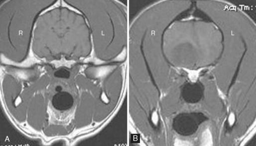
 MAGNETIC RESONANCE IMAGING SCAN A-B, T1-weighted magnetic resonance images from a normal (A) and abnormal (B) canine brain. Images are from rostral to midcerebrum. In MRI, the imaging characteristics of tissues are determined by the imaging sequence used. To define the tissue characteristics of a lesion, multiple different imaging sequences are performed, and the appearance of the lesion in these images is compared. The term intensity is used for describing the appearance of tissues in MRI.
PROCEDURE
< div class='tao-gold-member'>
Only gold members can continue reading. Log In or Register to continue
Stay updated, free articles. Join our Telegram channel

Full access? Get Clinical Tree


