Lumbosacral and Pelvic Injuries in Sports and Pleasure Horses

Chapter 50Lumbosacral and Pelvic Injuries in Sports and Pleasure Horses


Anatomical Considerations


Detailed anatomy of the ilium, ischium, pubis, sacrum, coxofemoral joint, sacroiliac joints, nerves, and major vessels is described elsewhere (see Chapters 49, page 564, and 51, page 583). The lumbosacral joint comprises five separate joints: the intercentral joint between the caudal aspect of the vertebral body of the sixth lumbar vertebra and the sacrum, between which is an intervertebral disk; two intertransverse joints; and two synovial intervertebral articulations between the articular processes of the sixth lumbar vertebra and the cranial articular processes of the sacrum. Movement is principally restricted to flexion and extension because of the large transverse processes.1 Congenital variations in anatomy can be seen, including fusion of the fifth and sixth lumbar vertebrae or sacralization of the sixth lumbar vertebra resulting in lumbosacral ankylosis. These result in stress concentration on adjacent joints. The biomechanical stresses of movement of the lumbosacral joint place particular compression and traction forces on the intervertebral disk, which may predispose to disk degeneration.


Clinical Signs


Clinical Examination


Clinical assessment of individual structures of the pelvic region by visual examination and palpation is not easy, especially in Warmblood and draft breeds, because of the large muscle mass of the hindquarters. Frequently, only the tubera coxae and tubera sacrale can be palpated. Large muscle mass may prohibit palpation of the greater trochanter of the femur. Atrophy of the hindquarter musculature is nonspecific and can reflect disuse because of pain arising anywhere in the limb, although atrophy of the muscles around the tail head often reflects injury to the tuber ischium or local nerve damage. Asymmetry of the height of the tubera sacrale is a common finding in horses in full work, free from lameness, although it may be seen along with poor performance or alterations in hindlimb gait. Apparent asymmetry may actually reflect differences in size of the dorsal sacroiliac ligaments. Alteration in muscle mass in the proximity of the tubera sacrale superficially can give a false impression of asymmetry of the tubera sacrale. Asymmetry of the tubera coxae may reflect a previous injury unassociated with ongoing pain. Poor muscle development in the lumbar region and over the hindquarters may make the tubera sacrale and the summits of the dorsal spinous processes of the lumbar vertebrae appear abnormally prominent. This should alert the clinician to the possibility of thoracolumbar or pelvic pain; however, this finding is nonspecific and may reflect the horse’s work history.


The pelvic region should be appraised visually and palpated systematically, and although preliminary assessment is usually best performed in the stable, for accurate evaluation of symmetry of the musculature and bony elements of the pelvic region the horse should be standing completely squarely behind on a firm, level surface with the horse looking straight ahead. In a horse with severe lameness this may not be possible because the horse may be unwilling to load the lame limb fully. Careful differentiation should be made between muscular and bony asymmetry. Muscle atrophy can make accurate assessment of symmetry of the pelvic bones difficult. To evaluate accurately the levelness of the tubera coxae, two assistants each must place an index finger on the craniodorsal aspect of each tuber coxae and extend the finger horizontally, or the tubera coxae should be marked using tape. Elevation of the tail may be necessary to identify muscle atrophy around the tail head, which may be seen along with nerve damage, or injuries of the ipsilateral tuber ischium.


Assessment of symmetry when the horse is unwilling to bear weight evenly on both hindlimbs is not easy, but particular attention should be paid to the way in which the limb is positioned. An abnormally straight limb may reflect luxation of the coxofemoral joint and secondary upward fixation of the patella. The greater trochanter of the femur of the lame limb may appear higher than that of the contralateral limb.


The muscles of the lumbar and pelvic regions should be assessed carefully to identify any area of abnormal muscle tension, pain on palpation, or unusual firmness. Firm stroking of the muscles first with a finger and then with a blunt-ended object (e.g., artery forceps) is useful to determine whether muscle spasm or muscle fasciculation is induced. Palpation of the caudal muscles of the thigh is also important because abnormal pain or tension can reflect primary muscle injury or an injury of the ipsilateral tuber ischium.


Firm pressure should be applied to the bony prominences to see whether pain or an abnormal reaction, such as sinking on the hindlimbs when pressure is applied to the tubera sacrale, can be induced. Both tubera coxae should be grasped simultaneously and the horse rocked from side to side to determine whether crepitus can be detected by palpation or auscultation, bearing in mind that the absence of crepitus does not preclude a fracture. Pull the tail while holding one hand over the coxofemoral joint; palpable crepitus may reflect a fracture, severe osteoarthritis (OA), or subluxation of the joint.


Careful, systematic examination of the pelvic canal region per rectum is also indicated to assess the aorta and iliac arteries, psoas musculature, the lumbosacral joint, the caudal aspect of the ilial shaft, and the pubis and ischium.


Pelvic injuries should also be considered when a clinician examines a recumbent horse that has fallen over a fence. Palpation of the pelvic region is even more difficult in these circumstances. Even with a severe fracture, palpating any abnormality may be impossible. The clinician should bear in mind that in the acute phase, local reflexes such as the patellar reflex and the withdrawal reflex may be suppressed, which does not necessarily reflect a spinal cord injury. Major fractures may be associated with rupture of one or more large vessels, resulting in potentially fatal internal hemorrhage. Thus it is important to assess the recumbent horse as a whole, monitoring pulse rate, color of mucous membranes, and capillary refill time. Increased pulse rate and progressive pallor of the mucous membranes are good indicators of major vessel rupture, such as laceration of the iliac artery after fracture of the ilial shaft.


Manipulation of the limb may be resented if pain is associated with the coxofemoral joint, but generally the responses to flexion of the limb, protraction, retraction, and abduction are rather nonspecific. A horse with pain associated with the sacroiliac joints or a coxofemoral joint may be reluctant to stand on one limb, with the other limb raised, and may behave awkwardly in anticipation of discomfort. However, the reaction is nonspecific, and one must bear in mind that some horses present difficulties in picking up the hindlimbs in the absence of any sign of lameness or poor performance. Difficulties in picking up hindlimbs may be caused by reluctance to accentuate weight bearing on the lamest limb, reluctance to flex the lame limb, or may be psychological. If the horse is a shiverer, unilaterally or bilaterally, the response to hindlimb flexion can be difficult to assess.


The degree and character of lameness depend on the underlying cause. Fracture or luxation of the coxofemoral joint results in acute-onset severe lameness. Lameness associated with other lesions in the pelvic region may vary in degree, not only among horses with similar lesions but also within and between examination periods. Pain from the coxofemoral joint frequently results in the horse moving on three tracks, with the nonlame limb being placed between the two forelimbs. On the lunge the horse may be inclined to break to canter rather than move with adequate hindlimb impulsion, but this is not specific for pelvic pain and is typical of many horses with hindlimb lameness. Pain associated with the coxofemoral joint or the greater trochanter of the femur sometimes results in the horse carrying the lame limb in canter. Lesions associated with the sacroiliac joints frequently result in the horse crossing over each hindlimb at the trot (i.e., plaiting), but this is not pathognomonic, and some horses move with a base-wide hindlimb gaitimage.2 The horse may move with reduced hindlimb impulsion rather than overt lameness. Although acute fractures of the tuber ischium invariably cause lameness, chronic injuries may result in loss of performance (e.g., jumping to the right) rather than overt lameness.


The response to flexion tests is nonspecific. The clinician should bear in mind that increased weight bearing on one limb, caused by flexing the contralateral limb, may accentuate lameness in the weight-bearing limb. Turning the horse in small circles, inducing rotational forces on the coxofemoral joint, may accentuate lameness associated with the coxofemoral joint.


Ridden exercise is invaluable in horses with a history of poor performance, reduced hindlimb impulsion, or low-grade lameness because frequently the lameness or restriction in hindlimb gait is accentuated. This may be most obvious in deep footing. Some horses with sacroiliac joint region pain or lumbosacral region pain show extreme reluctance to go forward freely. Affected horses may feel to the rider much worse than they appear to a trained observer. However, care must be taken to differentiate these horses from those with bilateral hindlimb lameness, thoracolumbar pain, or recurrent low-grade exertional rhabdomyolysis and those performing poorly because of the rider (see Chapter 97), previous poor schooling, or a combination of boredom and an unwilling temperament.


Analgesic Techniques


In horses with chronic lameness, reduced hindlimb impulsion, or poor performance, excluding the distal aspect of the limb as a source of pain by performing perineural analgesia of the fibular and tibial nerves and intraarticular analgesia of the three compartments of the stifle joint may first be necessary. If the response is negative, intraarticular analgesia of the coxofemoral joint may be indicated. This is relatively straightforward to perform if the horse is not well muscled and the greater trochanter of the femur is readily palpable. However, in the majority of heavily muscled, mature competition horses, needle placement must be guided by ultrasonography.4 Even if the needle is accurately positioned, retrieval of synovial fluid may be difficult. Extraarticular deposition of local anesthetic solution may result in transient paralysis of the obturator nerve and instability of the limb. The technique is described in Chapter 10.


Intraarticular injection of the sacroiliac joint cannot be achieved; however, infiltration of local anesthetic solution around the sacroiliac joint region may result in dramatic clinical improvement, presumably by alleviation of pain associated with the joint and periarticular structures. It cannot be considered an entirely specific technique and potentially could also influence pain from the lumbosacral joint and local nerve roots. The techniques are described on page 589. Infiltration of local anesthetic solution around the sacroiliac joint regions resulted in significant improvement in 95 of 108 horses with clinical signs suggestive of sacroiliac joint pain.5 Horses were reassessed ridden 15 minutes after injection. If local anesthetic solution is placed too far caudally there is the possibility of inducing sciatic nerve paralysis either unilaterally or bilaterally. In my experience, this is extremely rare (<2%), but if bilateral, the horse will become recumbent and may remain so for up to 3 hours before returning to normality.5


Diagnostic Imaging


Radiography


Radiographic examination of the pelvic region of a horse, anesthetized and positioned in dorsal recumbency or in the standing position, is described in depth elsewhere.6 Since the advent of nuclear scintigraphy and diagnostic ultrasonography the indications for radiographic examination have decreased. If the source of pain has been localized to the coxofemoral joint, radiographic examination is indicated to determine the nature of the pathological condition and hence prognosis. High-quality radiographs can only be obtained with the horse positioned in dorsal recumbency under general anesthesia. Evaluation of the sacroiliac joints can be difficult because of the superimposition of abdominal viscera. Identification of new bone formation on the caudal aspect of the joint and irregular joint space width are poor prognostic indicators. Nuclear scintigraphic examination gives accurate information about bone turnover, but anatomical detail is less well defined. Therefore radiography of a horse with a suspected acetabular fracture may be indicated at least 6 weeks after the onset of lameness to determine whether a suspected fracture involves the coxofemoral joint, which merits an extremely guarded prognosis for return to full athletic function in a mature horse.


Radiographs of the coxofemoral joint, caudal aspect of the ilial shaft, and the ischium obtained in the standing position may be satisfactory for confirmation of luxation or major fractures of the joint.6 A dorsal 50° proximal medial–ventrodistal lateral oblique image can be used to assess the integrity of the tubera coxae in a standing horse.7


Nuclear Scintigraphy


Nuclear scintigraphic evaluation of the pelvic region is useful for identifying fractures, stress reactions in bone, increased bone modeling associated with OA and other bony lesions, and evidence of rhabdomyolysis; for evaluating blood flow in the aorta, iliac, and femoral arteries; and for assessing the sacroiliac joints.10 Sensitivity of the technique in part depends on the angle of the gamma camera to the area of interest and the degree of overlying muscle mass. It is important to recognize that some lesions may be bilateral; therefore recognition of the normal scintigraphic appearance of the region in horses of different ages and different disciplines is important. The clinician should bear in mind that superficial bony structures such as the tubera coxae and tubera sacrale always have apparently greater radiopharmaceutical uptake (RU) than deeper structures. Unilateral muscle atrophy and shape of the pelvis may also confound interpretation.10-14


Radioactive urine in the bladder may complicate interpretation; therefore the judicious use of furosemide to induce urination before examination is essential.


Complete evaluation of the pelvic region requires dorsal images of the sacroiliac joints, oblique images of the ilial wings, caudodorsal and caudal images of the tubera ischii, and lateral and caudolateral images of the coxofemoral joints. Care must be taken in interpreting the appearance of the sacroiliac joints because age-related changes occur in normal horses.10-15 Swaying movement of the horse during image acquisition can result in images that mimic abnormalities, and using motion correction software is invaluable.


Differential Diagnosis


Fractures


Clinical features, diagnosis, and treatment of fractures of the pelvis in the young Thoroughbred (TB) racehorse have been dealt with in depth (see Chapter 49), and this section focuses on differences in mature athletic horses. The incidence of stress or fatigue fractures of the pelvic region in the mature horse is low, except in horses that race over fences, which have a substantial incidence of ilial stress fractures. The clinical features are similar to those in the young racehorse (see Chapter 49), although there is a higher prevalence of horses with fractures extending into the ilial shaft that have a guarded prognosis. The majority of other fractures result from external trauma.


Tuber Ischium


Trauma to or fractures of the tubera ischii sometimes occur in event horses that fall when jumping up onto a bank, may also occur in any horses as a result of a fall on the flat, and also have been recognized in horses from other disciplines with no known history of trauma.17-19 Lameness is usually acute in onset and severe. Mild localized swelling is easily overlooked unless the tuber ischium is suspected as a site of injury. The ipsilateral semimembranosus and semitendinosus muscles are usually sore to palpation. Atrophy of the muscles around the tail head often develops within 7 to 10 days. Usually crepitus is not palpable. In horses with chronic lameness, pain on palpation may not be evident, although the tubera ischii may appear asymmetrical, and the lameness may be only mild or moderate.


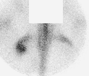
Diagnosis of a fracture of the tuber ischium can be confirmed using nuclear scintigraphy (Figure 50-1). Dorsal oblique and caudal images are useful. Usually increased radiopharmaceutical uptake (IRU) and an abnormal pattern of uptake are apparent.16 In some horses determining whether the fracture is complete and whether it has become substantially displaced may be possible. It is important to recognize that IRU may persist for many years after injury, and therefore the results of scintigraphic examination must be carefully correlated with the results of clinical examination.19 Discontinuity of the bone outline may also be confirmed using diagnostic ultrasonography. Limited radiographic examination can be performed in a standing horse, but it is most easily and safely done with the horse under general anesthesia. Less commonly there is entheseous reaction with IRU in both the tuber ischium and the ipsilateral semimembranosus and/or semitendinosus muscles. Ultrasonographic examination may demonstrate irregularity of the tuber ischium and decreased or increased echogenicity of the injured muscles depending on the acuteness or chronicity of injury.


Stay updated, free articles. Join our Telegram channel

Jun 4, 2016 | Posted by in EQUINE MEDICINE | Comments Off on Lumbosacral and Pelvic Injuries in Sports and Pleasure Horses

Full access? Get Clinical Tree

Get Clinical Tree app for offline access