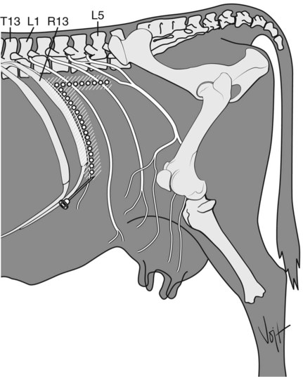
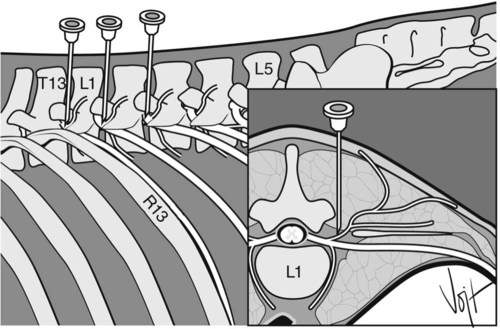
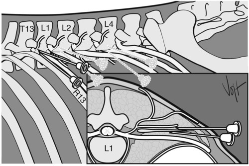
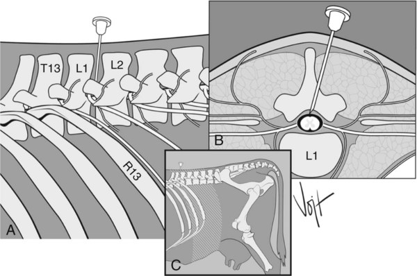
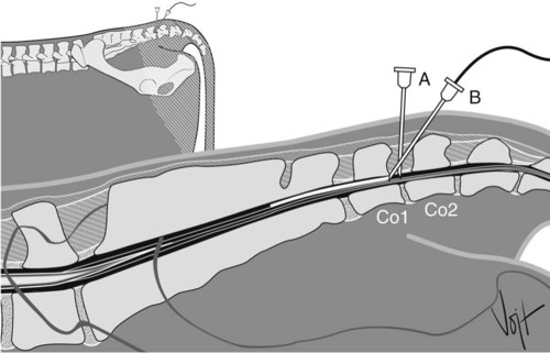
I Four techniques for producing local anesthesia of the paralumbar fossa in ruminants: II Abdominal surgeries in which these anesthetic techniques may be used: 1. Area blocked: skin and muscle layers of the flank and parietal peritoneum along the line of incision 2. Needle: 18-gauge, 7.6- to 10.2-cm 3. Anesthetic: 10 to 100 mL of 2% lidocaine 4. Method: make multiple subcutaneous injections of 0.5 to 1.0 mL of anesthetic, 1 to 2 cm apart with a 20-gauge, 2.54-cm needle; then infiltrate the muscle layers and parietal peritoneum through the desensitized skin. b. Use of routinely sized needles (2.5-cm, 20-gauge or smaller for skin block; 7.6- to 10.2-cm, 18-gauge for infiltrating the muscle layers and peritoneum). c. Incomplete block of deeper layers of the abdominal wall d. Formation of hematomas along the incision line e. Increased cost because of larger amounts of anesthetic used and time required 1. Area blocked: flank caudal and ventral to site of injection 2. Site: a line along the caudal border of the last rib and along a line ventral to the lumbar transverse processes from the last rib to the fourth lumbar vertebra (inverted L) 4. Anesthetic: up to 100 mL of 2% lidocaine in adult cattle, evenly distributed 5. Method: inject drug into the tissues bordering the dorsocaudal aspect of the last rib and ventrolateral aspect of the lumbar transverse processes, creating a wall of anesthetic enclosing the incision site b. Absence of anesthetic agent from the incision line minimizes edema, hematoma, and possible interference with healing a. Large volume of anesthetic required b. Length of time required to infiltrate such a long line c. Incomplete block of the deep layers of the abdominal wall (particularly the peritoneum) A Proximal paravertebral anesthesia (Farquharson, Hall, or Cambridge technique) 1. Area blocked: flank of side on which technique is performed 2. Nerves blocked: dorsal and ventral branches of T13, L1, and L2 and occasionally L3 and L4 (desensitization of L3 and L4 produce analgesia of the caudal-most part of the paralumbar fossa for cesarean section or ipsilateral foreteat and mammary gland; if L3 and L4 are blocked, the animal may become unable to stand) 3. Site: 2.5 to 5 cm from midline (Fig. 5-2); T13 immediately in front of transverse process of L1; L1 immediately in front of transverse process of L2; L2 immediately in front of transverse process of L3 4. Needle: 14-gauge, 1.3-cm needle, creating passage for a 16- or 18-gauge, 3.81- to 15.2-cm needle 5. Anesthetic: 20 mL of 2% lidocaine at each site 6. Method: the skin overlying the spinal column on the side to be desensitized is clipped, surgically scrubbed, and disinfected; palpate the lumbar transverse processes, starting from L5 and moving forward; L1 may be difficult to feel; measure 5 cm from midline; palpate the lumbar dorsal processes; injection site is at a 90-degree angle to the spaces between the dorsal processes; pass the needle vertically down until it hits the cranial edge of the transverse process and proceed down through the intertransverse ligament; inject 10 to 15 mL of 2% lidocaine below the ligament to block the ventral branch of the nerve (there should be minimal resistance to injection); withdraw the needle 1 to 2.5 cm sufficiently to inject 5 mL of 2% lidocaine above ligament, level with dorsal surface of transverse process to block the dorsal branch (resistance to injection); if the first lumbar transverse process cannot be palpated, anesthetize the other nerves first and then measure the distance between injection sites to find the site for blocking nerve T13 7. In sheep and goats, T13, L1, and L2 are desensitized in a fashion similar to the cattle method, but 2.5 to 3 cm off the midline and with less anesthetic (2 to 3 mL per site) 8. Advantages over local block a. Anesthesia of skin, musculature, and peritoneum; wide and uniform area of analgesia and muscle relaxation b. No additional restraint required c. Large quantities of local anesthetic not required d. Shorter postsurgical convalescent period; incision site avoided a. Procedure difficult in fat cattle and some beef cattle b. Arching of the spine caused by paralysis of back muscles c. No anesthesia of abdominal viscera d. Bowing out toward the area of incision (after unilateral blockade), making the closure of the incision more difficult B Distal paravertebral anesthesia (Magda, Cakala, or Cornell technique) 1. Area blocked: flank of side on which technique is performed 2. Nerves blocked: dorsal and ventral rami of T13, L1, and L2 3. Site: distal ends of lumbar transverse processes of L1, L2, and L4 (Fig. 5-3) 5. Anesthetic: 10 to 20 mL of 2% lidocaine at each site 6. Method: the skin overlying the spinal column on the side to be desensitized is clipped, surgically scrubbed, and disinfected; insert the needle ventral to the tips of the respective transverse process; inject anesthetic (up to 20 mL) in a fan-shaped infiltration pattern; withdraw the needle a short distance, reinsert it dorsal and caudal to the transverse process, and inject approximately 5 mL of the anesthetic. 7. Advantages of distal paravertebral nerve block over proximal paravertebral block a. Larger volume of anesthetic b. Variations in efficacy, particularly if the nerves follow a variable anatomic pathway C Segmental dorsolumbar epidural block (Arthur block) 1. Area blocked: the skin area caudal to the T13 or L1 spinous process and flank on both sides 2. Nerves blocked: T13 and anterior lumbar nerves, depending on the total dose administered 3. Site: epidural space between L1 and L2 vertebrae (Fig. 5-4) 4. Needle: spinal, preferably 18-gauge, 12.7-cm 5. Anesthetic: 8 mL of 2% lidocaine in a 500-kg cow, no more than 1 mL/50 kg of 2% lidocaine in sheep and goats 6. Method: the skin overlying the spinal column is clipped, surgically scrubbed, and disinfected; to reach the epidural space, insert the spinal needle 8 to 12 cm ventral and cranial at an angle of 10 to 15 degrees from vertical; piercing of the interarcuate ligament is felt as slight resistance during the insertion process; no blood or cerebrospinal fluid (CSF) can be aspirated, and also no resistance to the injection of anesthetic results after correct needle placement; if bleeding occurs, the stylet is placed into the needle and the needle is withdrawn after 2 to 3 minutes 7. Advantages over proximal or distal paravertebral anesthesia b. Small quantity of anesthetic c. Uniform anesthesia and relaxation of the skin, musculature, and peritoneum (begins 10 to 20 minutes after administration and continues for 45 to 120 minutes) I Caudal epidural anesthesia and desensitization of the internal pudendal nerve are commonly used in ruminants for obstetric manipulations, caudal surgical procedures, and as an adjunct treatment for control of rectal tenesmus; these techniques are not effective in pigs. A Low posterior or caudal epidural anesthesia 1. Area blocked: anus, perineum, vulva, and vagina 2. Nerves blocked: coccygeal and posterior sacral nerves 3. Site: first intercoccygeal space (Co1 to Co2: Fig. 5-5, A; more commonly and easily) or sacrococcygeal space (S5 to Co1) 4. Needle: 18-gauge, 3.8- to 5.1-cm (average dairy cow) 5. Anesthetic: 5 to 6 mL of 2% lidocaine or other drugs (Table 4-2) 6. Method: the skin overlying the spinal column is clipped, surgically scrubbed, and disinfected; locate the sacrococcygeal joint by moving the tail up and down; this joint moves very little and is located just anterior to the anal folds; the first intercoccygeal joint is easily located by its movement; it is much wider and is posterior to the anal folds; insert the needle exactly at the midline of the first intercoccygeal space at a right angle (approximately 10 degrees to vertical) to the skin surface; push the needle ventrally through the interarcuate ligament to the floor of the neural canal, which is at approximately 2 to 4 cm; withdraw the needle slightly (about 0.5 cm) into epidural space and test by injecting 1 mL of air; no resistance should be felt a. Minimal effect on cardiovascular and respiratory systems b. Little effect on organ systems c. Little problem with toxicity e. Good postoperative analgesia
Local Anesthesia in Ruminants and Pigs
Overview
Ruminants: Local Anesthesia for Standing Laparotomy
Anesthesia for Obstetric Procedures and Relief of Rectal Tenesmus
![]()
Stay updated, free articles. Join our Telegram channel

Full access? Get Clinical Tree