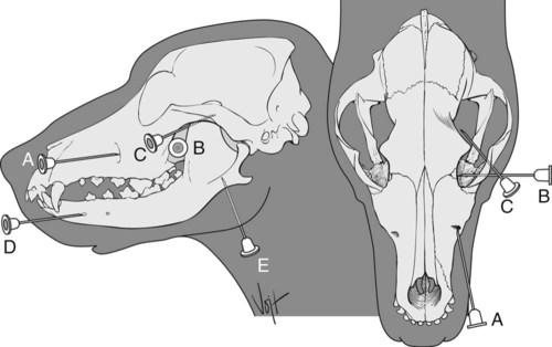
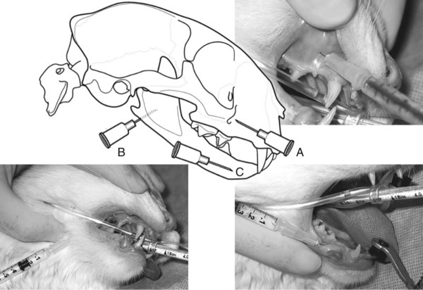
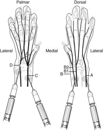
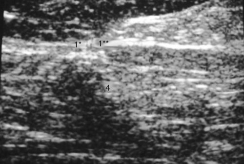
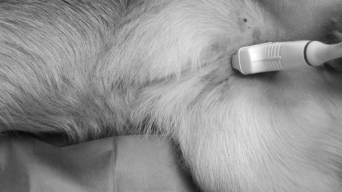
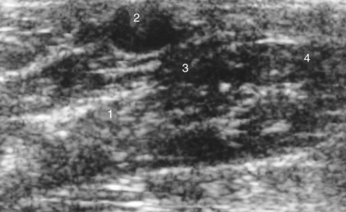
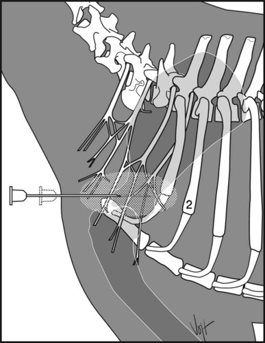
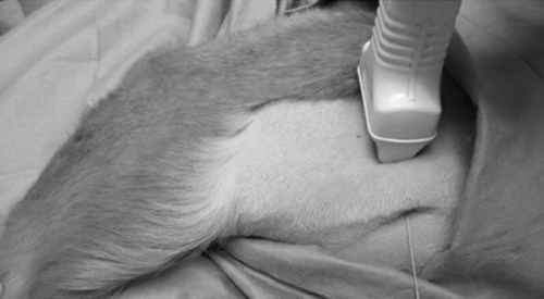
I Anesthesia of the upper lip and nose A Area blocked: upper lip and nose, roof of nasal cavity, and related skin ventral to the infraorbital foramen C Site: point of emergence of the nerve from the infraorbital canal (Figs. 7-1, A, and 7-2, A) D Needle: 22- to 25-gauge, 2.5- to 5-cm E Anesthetic: Use 1-2 ml of the chosen anesthetic or anesthetic combination. Do not exceed 1.5 mg of lidocaine/kg of body weight with or without 1.5 mg of bupivacaine or ropivacaine/kg F Method: insert the needle either intraorally or extraorally about 1 cm cranial to the bony lip of the infraorbital foramen; advance the needle to the infraorbital foramen, which can be felt between the dorsal border of the zygomatic process and the gingiva of the canine tooth II Anesthesia of maxilla, upper teeth, nose, and upper lip A Area blocked: maxilla, upper teeth, nose, and upper lip C Site: perpendicular portion of the palatine bone between the maxillary foramen and foramen rotundum (Figs. 7-1, B, and 7-2, B) D Needle: 22- to 25-gauge, 2.5- to 5-cm E Anesthetic: 1-2 ml of the chosen anesthetic or anesthetic combination. Do not exceed 1.5 mg of lidocaine/kg with or without 1.5 mg of bupivacaine or ropivacaine/kg F Method: insert the needle through the skin at a 90-degree angle, in a medial direction, ventral to the border of the zygomatic process and about 0.5 cm caudal to the lateral canthus of the eye; advance the needle in close proximity to the pterygopalatine fossa; local anesthetic is administered where the maxillary nerve courses perpendicular to the palatine bone between the maxillary foramen and foramen rotundum A Area blocked: eye, orbit, conjunctiva, eyelids, and forehead skin B Nerves blocked: lacrimal, zygomatic, and ophthalmic (i.e., ophthalmic division of the trigeminal nerve) C Site: at the orbital fissure (Figs. 7-1, C, and 7-2, C) D Needle: 22- to 25-gauge, 2.5-cm E Anesthetic: 1-2 ml of the chosen anesthetic or anesthetic combination . Do not exceed 1.5 mg of lidocaine/kg with or without 1.5 mg of bupivacaine or ropivacaine/kg F Method: insert the needle ventral to the border of the zygomatic process at the lateral canthus of the eye; the needle point should be about 0.5 cm cranial to the anterior border of the vertical portion of the ramus of the mandible; advance the needle medial to the ramus of the mandible in a mediodorsal and somewhat caudal direction until it reaches the orbital fissure IV Anesthesia of the lower lip C Site: rostral to the mental foramen (Figs. 7-1, D, and 7-2, D) D Needle: 22- to 25-gauge, 2.5-cm E Anesthetic: 0.5-1 ml of the chosen anesthetic or anesthetic combination. Do not exceed 1.5 mg of lidocaine/kg with or without 1.5 mg of bupivacaine or ropivacaine/kg F Method: insert the needle over the mental nerve, rostral to the middle mental foramen at the level of the lower second premolar tooth V Anesthesia of the mandible and lower teeth A Area blocked: cheek teeth, canine, incisors, skin, and mucosa of the chin and lower lip B Nerve blocked: inferior alveolar branch of the mandibular nerve C Site: point of entry of the nerve into the mandibular canal at the mandibular foramen (Figs. 7-1, D, and 7-2, C) D Needle used: 22- to 25-gauge, 2.5-cm E Anesthetic: 1-2 ml of the chosen anesthetic or anesthetic combination. Do not exceed 1.5 mg of lidocaine/kg with or without 1.5 mg of bupivacaine or ropivacaine/kg F Method: insert the needle at the lower angle of the jaw about 0.5 cm rostral to the angular process; advance the needle 1.5 cm dorsally against the medial surface of the ramus of the mandible to the palpable lip of the mandibular foramen I Anesthesia of the foot may be induced by the following techniques: A Infiltration of the tissues around the limb with local anesthetic solution (ring block) (Fig. 7-3) B Infiltration of the brachial plexus by local anesthetic (brachial plexus block) C IV injection of anesthetic into an accessible superficial vein in a distal extremity that is isolated from circulation by placing a tourniquet on the animal’s leg (IV regional anesthesia) D Injection of local anesthetic into the lumbosacral epidural space (anesthesia of the hind legs) E Perineural infiltration of sensory nerves in the limbs (nerve block) I Area blocked: distal foot, up to the elbow region II Nerves blocked: radial, median, ulnar, musculocutaneous, and axillary nerves III Site: medial to the shoulder joint (Fig. 7-4) IV Needle: 20- to 22-gauge, 7.5-cm (3.7-cm in cats); a spinal needle or catheter stylet works well V Anesthetic: 5- 10 ml of the chosen anesthetic or anesthetic combination. Do not exceed 1.5 mg of lidocaine/kg with or without 1.5 mg of bupivacaine or ropivacaine/kg A Insert the needle medial to the shoulder joint toward the costochondral junction and parallel to the vertebral column B Inject the anesthetic slowly as the needle is withdrawn; anesthesia can be obtained within 20 minutes and for up to 2 hours (total recovery requires about 6 hours) C Alternatively, use the same needle placement technique but use a nerve locator and insulated needle 1. The electrodes for the nerve locator controller are connected to the proximal end of the needle (uninsulated area) and the lateral thorax 2. Insert the needle with the nerve locator in the “On” position 3. As the needle gets closer to the brachial plexus, the paw will begin to twitch. The goal is to have a maximum twitch with minimal amperage on the controller. 4. When a maximal twitch is achieved, inject local anesthetic to obliterate the twitch 5. Place the needle at a slightly different angle with the goal of obliterating all twitches in the foot. This will give a 100% block. 1. Relatively simple and safe to perform 2. Produces selective anesthesia and relaxation of the limb distal to the elbow joint 1. Relatively long waiting period (15 to 30 minutes) required 2. Occasional failure to obtain complete anesthesia, particularly in overweight dogs. This failure is minimized when a nerve locator is utilized. I Area blocked: The femoral nerve innervates the cranial muscles of the thigh. All remaining muscles of the limb are innervated by the sciatic nerve. II Site: The femoral nerve is imaged at the medial aspect of the thigh. The sciatic nerve is imaged just distal and caudal to the greater trochanter. III Needle: 22-gauge, 2.5 to 3.7 cm IV Anesthetic: 2-4 mL (total) of the chosen anesthetic or anesthetic combination. Do not exceed 1.5 mg of lidocaine/kg with or without 1.5 mg of bupivacaine or ropivacaine/kg A Clip the hair from the sacroiliac region just below the stifle on the dorsal, lateral, and medial aspects of the limb B Clean the skin and apply coupling gel C For the sciatic nerve block, place the dog in lateral recumbency 1. Place the ultrasonic transducer on the transverse plane just distal and caudal to the greater trochanter and then direct it towards the distal aspect of the thigh (Figs. 7-5 and 7-6) 2. The sciatic nerve is imaged from the dorsal aspect. Transverse images can also be obtained by positioning the transducer parallel to the dorsal anatomical plane caudal to the greater trochanter and cranial to the ischiatic tuberosity. Longitudinal images of the sciatic nerve can be obtained by rotating the transducer 90 degrees from the position used to obtain the transverse images. Several acoustic windows are available to approach this nerve along the lateral surface of the thigh. D Approach of the femoral nerve. This description uses a mid-femur approach. The femoral nerve is imaged at the medial aspect of the thigh. Position the dog in lateral recumbency. 1. Place the transducer in the inguinal skin crease cranial to the pectineus muscle while the contralateral limb remains abducted (Figs. 7-7 and 7-8). The nerve is scanned cranially to the pectineus muscle. This is the only acoustic window that allows a good approach to the femoral nerve. E Both the sciatic and femoral nerves are approached using the acoustic windows described earlier. The accuracy of the nerve location is confirmed by use of a peripheral nerve stimulator. 1. Connect an insulated needle to the nerve stimulator and insert along the plane of the ultrasound transducer. When the tip is seen close to the nerve, the nerve stimulator is turned on. 2. When the needle is close to the sciatic nerve, the tarsus should flex or extend. When the needle is close to the femoral nerve, the knee should extend. 3. Once the nerve is located via ultrasound and confirmed with the nerve–stimulator-induced twitch, inject local anesthetic incrementally to surround the entire nerve. This will look like a donut on the ultrasound image. Usually no more than 0.3 mL of local anesthetic is required to produce a block at each nerve.
Local Anesthesia in Dogs and Cats
Overview
Regional Anesthesia of the Head
Anesthesia of the Foot
Brachial Plexus Block
Ultrasound Guided Rear Limb Block (sciatic and femoral nerve blocks)
![]()
Stay updated, free articles. Join our Telegram channel

Full access? Get Clinical Tree