Chapter 68 Laparoscopic Surgery in the Elephant and Rhinoceros
Traditional abdominal surgery in the elephant and rhinoceros has rarely been performed and, in those cases in which it has, success has been limited.1,4 Emergency cesarean sections in elephants have been attempted and have proved fatal for both the calf and mother. Successful castration of bull elephants has been described.2,3,12 In these reports, young animals were castrated in a single procedure, whereas adults required multiple procedures. These elephant castration procedures were difficult and it was not uncommon for incisions to break down and heal by second intention. An abdominal exploratory and ovariectomy has been reported in an Indian one-horned rhinoceros, but the animal died 48 hours after the surgery.11
Abdominal surgery in the elephant and rhinoceros is difficult for several reasons, the first being the animal’s overall size and how it affects the surgeon’s ability to reach and manipulate organs. Second, there is considerable thickness to the skin and body wall, which requires larger incisions and an inordinate amount of time for closure. Surgical sites may also be predisposed to dehisce and break down.2,12 An additional complicating anatomic variation in elephants is their peritoneum. Unlike most other mammal species, the peritoneum is covered by a fibroelastic layer and is itself redundant and only loosely attached to the body wall.5 For this reason, entering the peritoneal cavity of elephants, even after making an incision through the dermis and associated muscle layers, may be very challenging. Finding a route to and through the peritoneum is much more of an issue when animals are in lateral recumbency (versus standing), when there is less tension on the peritoneum. Rigid laparoscopy, or minimally invasive surgery (MIS), offers a variety of advantages over traditional surgical procedures. In humans and animals, MIS has been shown to be less painful, requires less healing time, allows faster return to normal function, and has less chance for infection.19 For the elephant and rhinoceros, MIS has these advantages and thus makes abdominal surgery a realistic and much less risky procedure. Laparoscopic abdominal surgeries have been successfully performed in the African elephant, white rhinoceros, and black rhinoceros.6,13,15–17 Procedures have included abdominal exploration with diagnostic sampling and a variety of reproductive surgical techniques.
Laparoscopy in the Elephant
Animal Positioning
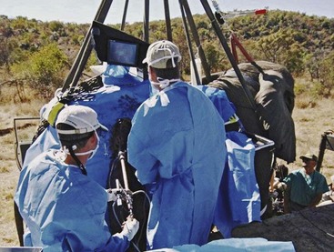
Abdominal laparoscopic surgery in elephants is best accomplished in a standing position, either with the animal sedated and in a restraint device or under general anesthesia, with the animal in a standing position while being suspended from a crane truck.6,15–17 Using MIS and maintaining the elephant in a standing position allows for small incisions, rapid access to the abdominal cavity, and excellent overall surgical success.7 In captive situations, in which sedation may be used along with some degree of manual restraint, abdominal laparoscopic surgery may be readily accomplished. We have successfully used butorphenol and detomidine in conjunction with local analgesic nerve blocks with the elephant in a restraint chute (Fig. 68-1). The patient is sedated to the level at which it cannot lift its trunk and maintains a wide stance on all four legs, but is not likely to lie down. Once sedated, a regional analgesic block is accomplished using a 5-inch, 18-gauge spinal needle. In elephants, an elevated platform is used to place the surgeons at the level of the paralumbar fossa. This standing sedation approach has been used for abdominal exploratory and reproductive procedures in both adult elephants and rhinoceroses.
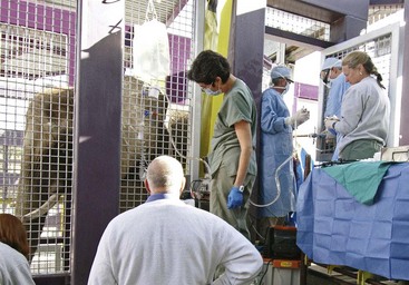
In free-ranging elephants, for which restraint facilities do not exist and the location of the patient is difficult to predict, the animals are placed under general anesthesia via a remote injection system. Once the patient is laterally recumbent, large padded straps or ropes (5-ton capacity with foam or wool padding) are placed around the proximal base of each leg and connected to the hook of a crane truck. These ropes are looped around the axilla or inguinal areas, ensuring limited pressure on the thoracic or abdominal cavities (Fig. 68-2). Limited pressure on the thorax greatly improves respiration and limited pressure on the abdominal cavity assists in insufflation and laparoscopic visibility. Once the ropes have been placed around each leg and secured to the crane, the elephant is lifted into a standing position. An additional rope is placed around the base of the tusks and attached with the other ropes so that the head is held in a normal upright position. In most cases, these animals are intubated to facilitate assisted ventilation when insufflation is applied.
Surgical Equipment
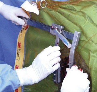
The nonlaparoscopic surgical instruments used for creating and closing the abdominal incisions are commonly available through veterinary surgical catalogues. In most cases, the large versions are used. The procedure benefits from two to four no. 8 surgical scalpel handles with no. 60 scalpel blades, and two to four long no. 4 scalpel handles with no. 22 scalpel blades. Elephant and rhinoceros skin is tough, dense, and thick, so many blades are generally used for a single incision. It is always better to have too many blades than not enough. Other instruments include penetrating towel clamps, large Mayo scissors, long needle holders, long thumb forceps, vulsellum forceps, wire needle holders, and wire cutters. Modified Finocetto rib spreaders are also used to distract the skin while making the primary incision (Fig. 68-3).
Laparoscopic Surgical Instruments
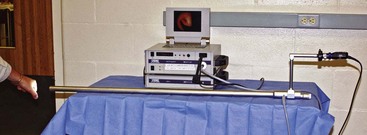
Laparoscopic surgical equipment for minimally invasive surgery in the megavertebrate species must be longer and stronger than traditional equipment. The telescope—a rigid laparoscope— should have at least an 80-cm working length for bull elephants younger than 20 years and for cow elephants. In larger bull elephants, a working length of 100 to 112 cm is helpful (Fig. 68-4). A traditional telescope may be used but, to reduce the number of portals necessary, an operating laparoscope is more useful. Specialty telescopes (Karl Storz Endoscopy, Tutlingen, Germany) with two light source input posts will improve the amount of light that may be delivered into a potentially large peritoneal space. Visibility may be difficult because of the size of the abdominal cavity and the amount of fat and bowel distension that may occur. Having an appropriate amount of light is important. In the rhinoceros, depending on the size of the animal and procedure being attempted, an equine telescope (Hopkins Telescope, Karl Storz Endoscopy, 10 mm × 57 cm, 30-degree angle) may be used.13
Stay updated, free articles. Join our Telegram channel

Full access? Get Clinical Tree


