Lameness in the Show Hunter and Show Jumper

Chapter 115Lameness in the Show Hunter and Show Jumper


Historical Perspective


Show jumping and related competitions have origins in hunting sport and military tradition. The show hunter, most popular in North America, has evolved from the traditions of fox and stag hunting. These horses were expected to provide fast, safe, and athletic passage for the rider, and considerable pride was taken in being well mounted and having the horse admired. Today in the show ring these horses are judged for beauty, athletic ability, manners, and way of going. Jumping style is important and must be coupled with consistent performance.


Some competitions encourage the development of a young horse to a higher level of training, others award a mature horse for outstanding performance, and others separate amateur and professional riders. Horses often are selected based on suitability for a particular division of competition.


The modern show jumper has many of its origins with military traditions. Many cavalry officers were by necessity highly skilled and accomplished horsemen. Thus when the era of the modern Olympics began, the equestrian competitors were military men. With the mechanization of the military and the replacement of the cavalry with motorized transport, the private sector became more involved in Olympic show jumping. Many of the early civilian competitors were retired military men. Show jumping has become increasingly more popular, and many talented riders have emerged on the national and international scenes. Since the 1960s many women have entered this sport, once dominated by men.


Structure of the Sport


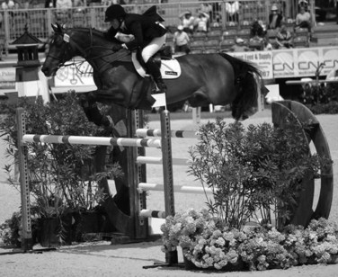
Show jumping combines athletic effort of the horse and rider. The scoring process is objective, with the winner jumping the course with the fewest rails knocked down and, in the jump off, in the fastest time. Heights of fences range from 1 m at novice level to 1.7 m for advanced competitions. The course should be completed within an allowed time, and some competitions are considered as speed classes in which jumping and time faults are combined. As the jumps get bigger, the potential for injury increases, and many conditions develop from repetitive strain. Many of the fences are set at distances to each other so that the horse must adjust stride length to fit in the appropriate number of strides. A good horse must have explosive power and great athleticism, combined with carefulness—a desire not to hit fences. Contrary to many equine sports, similar numbers of mares and geldings or stallions compete (Figure 115-1).


Some older horses compete at levels of competition lower than they have reached to be schoolmasters for less experienced riders. These older horses may experience unique problems related to age and use. Today show jumping is a highly diverse and competitive sport enjoyed all over the world from beginner level to the level of the Olympic Games. At the top level the sport is entirely professional, with horses changing hands for huge prices and with large amounts of prize money available, putting pressure on the veterinarian to keep horses sound. Competition continues throughout the year, with outdoor shows during the summer months and indoor shows during the winter, so horses potentially get few breaks.


Training


Training of the hunter and jumper emphasizes using the hindquarters for engagement and collection, which places more weight and stress on the hindlimbs as they are brought forward and under the rider during locomotion. Such a posture is somewhat unnatural for the horse, whose normal inclination is to distribute weight over the forehand. These stresses may contribute to or accelerate the development of problems of the thoracolumbar and pelvic regions, and ligaments and joints of the hindlimbs. Early in the training of a young horse, lameness often reflects musculotendonous problems and relates to lack of adaptation to work.


When training over fences begins, new loads on the hindlimbs occur that place more stress on soft tissues and joints. Stresses on the forelimbs also are increased because they are involved in takeoff and landing. The forelimbs are involved in setting up the jump and aiding in the directional change from horizontal to vertical. On landing the forelimbs receive considerable impact loads and absorb the entire weight of the horse. Increased load places more stress on the foot, distal limb joints, and the soft tissues of the forelimbs.


Horses must learn to adjust stride length by shortening or lengthening the stride, to jump from a line perpendicular to a fence or at an angle, to turn quickly, to change leading legs in canter, and to jump from variable speeds of approach. Unwillingness to change leads or always favoring one lead when landing after a fence may indicate a problem (see Chapter 97). Much of the basic flat work training is similar to dressage.


Many lameness conditions encountered early in a horse’s training begin as subtle performance-limiting problems, progress relatively slowly, and may disappear with further conditioning. However, excessive training leads to the development of important problems such as chronic muscle soreness, fatigue, joint inflammation, and behavioral changes. Slow, steady work with a gradual buildup in exercise intensity and duration results in fewer joint and soft tissue problems later.


Training and Competition Surfaces


Training, warm-up, and competition surfaces play a substantial role in the development of lameness in a jumper. Soft, deep footing requires much more effort by the horse and is responsible for early fatigue of muscles, tendons, and ligaments. Injuries include gluteal muscle strain and spasm, suspensory desmitis, desmitis of the accessory ligament of the deep digital flexor tendon (ALDDFT), and superficial digital flexor (SDF) tendonitis as well as inflammation of or trauma to any of the soft tissue structures in the hoof, pastern, and metacarpal and metatarsal regions. Sandy soils contribute to the development of hoof wall problems. Hard surfaces may result in bone- and joint-related injuries, as well as distal limb and foot problems such as subsolar bruising, distal interphalangeal (DIP) joint synovitis, and osteoarthritis (OA) of the proximal interphalangeal (PIP) and DIP joints.


Formerly most high level outdoor competitions in Europe took place on grass, but there is an increasing trend for some of the higher-level competitions to take place on all-weather surfaces. The nature of the footing depends highly on the weather and can vary extremely. To enhance traction, screw-in studs often are used in the front and hind shoes. Studs may be used in the medial and lateral branches of the shoe or just the lateral branch to reduce risk of interference injuries. If studs are used in only one branch of the shoe, this immediately creates mediolateral imbalance and the potential for abnormal torque. Studs also alter the dorsopalmar balance, especially if the ground is firm. Horses that flex the carpi excessively during jumping may have to wear a girth guard to protect the sternal region from self-inflicted injuries. If a horse is jumping on firm going, studs concentrate the forces of impact and can predispose the horse to deep-seated bruising of the foot. There is a tendency in horses with poor-quality horn for the hoof wall to break in the region where studs are placed. In some circumstances studs may halt the normal slide of a foot on landing and thereby predispose to injury.


Excessive work, such as long period of lunging a hunter to calm it down, prolonged competitive efforts, and long show schedules of repeated competitions play a role in the development of many injuries in hunters and jumpers.


Conformation and Lameness


Conformational abnormalities of the foot predispose horses to lameness. An underrun heel, long toe, and a broken hoof-pastern axis frequently contribute to palmar foot pain and DIP joint synovitis. Horses with improper medial-to-lateral hoof balance may develop a sheared heel, crushed bar, and chronic foot pain. Base-wide or base-narrow forelimb conformation may contribute to injuries to the suspensory ligament (SL), its branches, and the distal sesamoidean ligaments.


Toe-in or toe-out forelimb conformation may predispose horses to PIP and metacarpophalangeal joint problems. Short, more upright pasterns predispose horses to navicular disease and DIP joint and PIP joint synovitis or OA. These horses are often straight through the shoulder as well and lack the stride and extension necessary for jumping. Long, more sloping pasterns sometimes are associated with sesamoiditis and soft tissue injuries to the distal aspect of the limb.


Over-at-the-knee conformation may predispose horses to SL strain and should be avoided in selecting a jumper. Offset-knee conformation predisposes a jumper to medial splint problems. However, attention always should be paid to subclinical contralateral hindlimb lameness in a mature horse that develops an acutely painful medial splint bone. Subtle angular limb deformities are rarely a problem, provided proper attention is paid to shoeing and hoof balance.


Extremely sickle-hocked conformation is associated with weak hindlimbs and places more stress on the plantar tarsal soft tissues and the centrodistal and tarsometatarsal joints. A straighter hock is actually more desirable, but an overly straight hock may predispose to SL strain and distal hock joint pain, especially in association with an extended fetlock and long pastern conformation.


Horses that are extremely straight through the stifle are poor jumping prospects and have a high incidence of instability and upward fixation of the patella. A more angular stifle gives the horse a longer, more powerful stride and is thought to provide more strength for jumping.


A long, sloping hip and croup are desirable characteristics in a jumping horse, providing strength and power. Horses with a flat croup often suffer from thoracolumbar and sacroiliac pain. Asymmetry of the tubera sacrale and tubera coxae can be seen in the absence of lameness and may reflect previous trauma. An experienced rider usually can manage these horses.


Horses with excessively base-wide or base-narrow hindlimb conformation place abnormal stress on the feet and joints. Base-wide horses may have an increased incidence of hock problems, whereas base-narrow horses may have more stifle problems.


Lameness Examination


Before a horse is examined because of a suspected lameness problem, several factors should be considered. If the horse is currently competing, will any diagnostic tests or treatment have an effect on the horse’s ability to continue competition? Knowledge of the competition rules and drug use guidelines are essential. Has the horse recently competed? Has a change in exercise intensity or duration been made? Does the horse have a new trainer? When was the horse last shod? If the horse has been competing away from home, it is important to determine if another veterinary surgeon already has examined and treated the horse, and if so with what.


The lameness examination should begin as the horse is walked from the stall or paddock to the examination room or area, because movements such as a small circle to reverse its direction may offer clues as to which limb or limbs may be affected. It is not uncommon for there to be concurrent lameness in more than one limb. Careful observation and palpation of the joints of each limb as well as the neck and back are performed while the horse is held quietly. Passive flexion tests and evaluation of the lateral movements of the head, neck, and back are performed.


A dynamic examination should be performed beginning with an observation of the horse at the walk in a straight line, in small circles, and in a figure eight. The horse should next be examined at the trot in hand on a firm, level surface. Lunging on firm footing in small circles in both directions should be performed, as this often exacerbates subtle lameness, especially if involving the foot.


It is sometimes necessary to see the horse ridden to determine which limb is lame. The rider may also be asked to change the diagonal at the trot and figure eight to increase the load on a specific limb when it is on either the inside or the outside of the circle.


Examinations performed after strenuous exercise or after competition may be useful; more subtle lameness may then be more obvious. It should also be noted and recommended that the veterinarian observe the “way” or “style” the horse trains and jumps. Defects in the horse’s position and way of jumping that would have otherwise gone unnoticed may be observed during a routine physical or lameness examination. Watching videos of the horse may also allow the practitioner to see abnormalities of the horse’s balance or gaits when in motion. Knowing what is “normal” may also help the practitioner detect early or subtle changes in movement before an obvious lameness is manifest.


Proximal and distal limb flexion tests should be performed on all limbs. The method and duration of flexion are a matter of personal preference but should be consistent and interpreted with care, bearing in mind that joints and soft tissue structures may be stressed simultaneously. It has become common practice for top-level competition horses to be examined periodically throughout the year to try to detect early warning signs of impending problems. A positive response to flexion is often followed by treating the stressed joint(s). The true value of this practice is difficult to determine objectively.


Once the lame limb(s) has been identified, the limb is carefully reexamined. Hoof tester response should also be assessed. An obvious source of pain may be identified, but perineural or intraarticular analgesia often is required to localize the source(s) of pain. Results are sometimes confusing and always should be related to the clinical examination.


Aseptic preparation is essential for intrasynovial injections to minimize the risk of infection. Two authors (RPB, CBM) also administer gentamicin intravenously before entering any synovial space. The conditions of the work area and the temperament of the horse being examined influence which analgesic technique to use. Intrasynovial analgesia may be delayed or not performed if no clean, dry place is available for safe injection. Although intraarticular analgesia is considered to be more specific than perineural analgesia, it may influence periarticular pain. In some instances regional or intraarticular analgesia may not be safely performed without first tranquilizing the horse. This may interfere with the lameness examination, especially if the lameness is low grade. Lameness is reassessed after the effects of sedation have worn off. Alternatively the horse is sedated, the suspected joint is treated with the appropriate drug(s), and the horse is reevaluated in a few days. The treatment is therefore substituted for intraarticular analgesia. In situations in which subtle lameness makes interpretation of nerve blocks difficult, in those horses with multiple-limb lameness, in horses that are difficult or dangerous, or when comprehensive blocking fails to localize the lameness, other techniques such as nuclear scintigraphy should be considered.


When horses are competing regularly, especially away from home, owners, riders, trainers, and peers often put pressure on the veterinarian to treat the horse based on an index of clinical suspicion rather than on a complete lameness evaluation including local analgesia. Although this can be successful and a positive response to treatment clearly indicates a correct diagnosis, one must bear in mind that some injuries do require rest for the best long-term outcome. A merely transient response to treatment or a lack of response warrants further investigation of the lameness, and this routinely should include local analgesic techniques.


Failure to Make a Diagnosis


Every veterinarian, no matter how astute as a lameness diagnostician, eventually will be confused or unsure or simply have no idea as to why a particular horse is lame, and consultation with associates or referral to other experts should be considered. Just as a good relationship with a farrier is paramount to the successful management of many foot-related problems, good relationships with other veterinarians are necessary and may be helpful when one is faced with a difficult or confusing lameness. The veterinarian must be honest and open about the horse with the owners and trainers; inclusion of other professionals in the case or referral for advanced imaging may ultimately lead to both an accurate diagnosis and enhancement of the veterinarian’s relationship with the client if handled well. Reexamination at a later date also may be beneficial. Some bone lesions may take a few weeks to become visible radiologically, so the veterinarian should consider reimaging if the pain causing lameness has been localized to a specific area.


The use of many medications for a “shotgun” approach to treatment may be a veterinarian’s only hope when all diagnostic methods are either unrewarding or not available and if previously attempted “trial therapies” have failed. Such medications include but are not limited to nonsteroidal antiinflammatory drugs (NSAIDs), doxycycline, tiludronate, intramuscular polysulfated glycosaminoglycans (PSGAGs), intravenous hyaluronan, and oral nutraceuticals.


Diagnosis and Management of Common Causes of Lameness


Foot Pain


Foot Soreness


The most common site of forelimb lameness and overall lameness in a hunter or jumper is the foot. The horse naturally supports 60% to 65% of its body weight over the forelimbs, and impact forces during jumping dramatically increase load and structural stresses in tissues within the hoof capsule. This may be exacerbated by training regimens that do not focus on improving strength from behind and therefore the overall balance of the horse.


The manner in which a horse is shod has tremendous importance in the development of hoof- and foot-related problems. A long toe and underrun heel are common hoof conformational defects and frequently contribute to heel pain because of hoof wall separation or bruising in the heel, quarter, and bar areas. The heel itself may be excessively long and collapsed inward, and the horse actually may be bearing weight on the outer wall. This often results in sensitivity to hoof tester pressure applied to each heel bulb and when the heel bulbs are squeezed together. A flattened and chronically bruised heel and bar area (corns) may be seen after removal of the shoe.


Lameness is often improved by analgesia of the palmar digital nerves. Lateromedial radiographic images of the foot may reveal that the distal phalanx is abnormally oriented, in extreme situations so that the palmarmost aspect of the bone is lower (more distal) than the toe (so-called “negative palmar angle”). The so-called “negative palmar angle” appears to occur more commonly in the United States than in Europe. The solar margins of the distal phalanges may be irregular. In horses with chronic lameness, deep digital flexor (DDF) tendonitis and distal sesamoidean impar desmitis may contribute to pain associated with an underrun heel. However, these soft tissue injuries have been documented using MRI in show jumpers with both poor and good foot conformation (SJD).


Some horses with underrun heels and poor angulation of the distal phalanges do not respond well to shoeing. Removing the shoes, trimming back the abnormal heel wall, and placing the foot in a support bandage are recommended. One author’s (RPB) preference is to fashion a cushion support for the palmar aspect of the hoof using a two-part putty elastomer material (EDSS, Equine Distal Support System, Penrose, Colorado, United States). Using the sulci of the frog for support is believed to suspend the heel and promote the new growth to be more vertical in its orientation. This process, however, requires a long-term commitment by the owner and trainer, because new heel growth may take up to 6 months to be sufficient for the reapplication of shoes. One author floats the heel using a image rim pad and either a heart bar shoe or a pour-in pad in an effort to keep the horses in work and competition (CBM).


Attention should be paid to the role of training and overall management of the horse, specifically as it relates to hindlimb strength and comfort.


Subsolar Bruising


Horses with subsolar bruising often respond well initially to Epsom salt poultices or cooked linseed hoof packs and NSAIDs (e.g., phenylbutazone) followed by corrective shoeing. Rasping excessive toe from the solar surface proximally up the dorsal wall to create a 45-degree angle with the ground surface and application of a shoe fitted full at the heel may be of benefit by removing resistance to breakover. Ideal breakover is located between two points; the first is located by extending a line distally along the dorsal surface of the distal phalanx to the bearing surface, and the other by drawing a perpendicular line from the dorsal distal aspect of the distal phalanx to the bearing surface. Egg bar shoes may be required to gain adequate heel support. Shoes such as the EDSS natural balance shoe (Equine Digital Support System), with the web behind and squared off at the toe, also improve breakover and reduce stress in the palmar portion of the foot. We do not recommend the long-term application of plastic wedge pads or egg bar shoes because they actually may contribute to further crushing of the heel and promote the heel bulbs to slide forward and grow horizontally. Leather pads may be helpful in some horses if sole pain is present and sole protection is desired. In horses with a chronic problem, long-term use of aspirin (60 g once daily or 20 g bid for 5 days) may be helpful. Hoof growth supplements containing biotin and methionine also may be of benefit, and we recommend feeding of biotin (100 mg to 1 g for 3 to 12 months, either alone or combined with zinc, cysteine, and dl-methionine daily) to promote hoof wall growth. Careful attention should be paid to the condition of the gastric mucosa with long-term NSAID use in show horses, because gastric ulceration may occur or worsen. Concurrent administration of acid pump inhibitors such as omeprazole should be considered.


Extreme sensitivity to hoof testers may be evident along the periphery of the sole at the level of the distal phalanx. Such pain may be associated with bruising, solar margin fractures, laminar inflammation or inflammation of the distal phalanx caused by chronic concussion from hard ground, or excessive sole pressure from the shoe. These conditions may be more common in North America (with its harder and dryer footing) than in Europe. Radiology is necessary for diagnosis of solar margin fractures of the distal phalanx. Shoeing should be directed at reducing local pressure on the affected areas and improving the overall hoof balance. Egg bar shoes and rim pads are often effective, but soft sole pours that provide extra cushion and shock absorption also help. Two-part putty elastomer is thought to benefit by providing support and lift from the sulci of the frog. Care should be taken with a pour or putty elastomer to avoid overfilling, causing excessive sole pressure.


Subsolar Abscess


Subsolar abscesses occur commonly and result from shoe nails improperly applied, poor environmental conditions, a shoe moving slightly, and poor hoof structure. Onset of clinical signs may be rapid, such as immediately after a show, or may occur within the first several days after shoeing. The additional trauma of jumping exacerbates the condition, leading the rider or trainer to suspect trauma or serious injury. Warmth in the hoof wall, increased digital pulse amplitudes, and focal, extreme sensitivity to hoof tester application are usually diagnostic, provided that the hoof horn is not excessively hard. Perineural analgesia is rarely necessary to confirm the suspected diagnosis and also may be confusing, because not all horses respond positively. After blocking and trotting the horse to reassess lameness, reexamination of the solar surface of the foot may reveal purulent drainage from the area of suspicion. Treatment is directed toward liberal opening of the solar surface of the foot at the point of maximum sensitivity to establish adequate drainage. If drainage is not established, bandaging the foot with hyperosmotic agents such as products containing magnesium sulfate (Epsom salts) is recommended. Twice daily soaking of the foot with a hot, supersaturated solution of Epsom salts with the bandage left on is also recommended for 3 to 5 days. Once drainage has been established, the foot is bandaged in a similar fashion, and NSAIDs also may be administered to reduce the inflammation. Antibiotics rarely are indicated but are sometimes used if soft tissue swelling occurs above the coronary band.


Jun 4, 2016 | Posted by in EQUINE MEDICINE | Comments Off on Lameness in the Show Hunter and Show Jumper

Full access? Get Clinical Tree

Get Clinical Tree app for offline access