Lameness in the Polo Pony

Chapter 119Lameness in the Polo Pony


History of the Sport


Polo was the first equestrian sport in recorded history. With strong ties to military traditions, the game originated in China in ad 272 and was often substituted for war games in preparation for military battle. Polo evolved into an organized sport and spread into Greece, India, and China, where the British colonies adopted the game. During the nineteenth century, the game became more refined in Great Britain and eventually found its way into the northeastern United States in 1876. Today polo continues to be one of the fastest and most dangerous equine sports in the world. Polo is no longer a sport only for the wealthy; an increasing number of small clubs start up each year that attract people of moderate incomes to take lessons, buy horses, and begin to play. The sport has become more complex, with international professional players competing year-round on different teams. Thus professional polo coaches, umpires, trainers, and breeders have emerged, solidifying polo as a genuine equine sport industry. Playing seasons in the northern and southern hemispheres are followed by nomadic players, grooms, horses, and spectators. During the winter months in the United States, thousands of horses enter Florida and California, where the tropical climate is conducive to world-class polo tournaments. During the spring, summer, and fall seasons these horses travel across the Midwest, up the northeast coast and into Canada. The high-goal season begins in the summer in England and Spain, but in the southern hemisphere, the season in Argentina commences in the fall.


Polo as an Industry


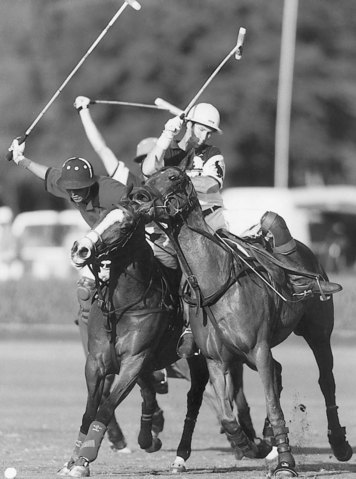
Today three types of polo are played: outdoor, indoor (arena), and snow polo. Outdoor polo is by far the most popular and is played on a large, finely manicured grass field measuring 274 m (300 yards) by 137 m (150 yards) (Figure 119-1). Injuries are related to fatigue (because of the distances covered), stopping, turning, speed, and surface consistency, depending whether the soil beneath the grass is compact or soft. Arena polo is played in much smaller arenas and is more common in collegiate settings. Injuries tend to be less frequent and are usually impact related. Snow polo is regarded as a novel exhibition sport played on the surface of a frozen lake and produces surprisingly few injuries. Obviously, footing and surface conditions often can be responsible for the type of lameness seen. Heavy, soft, grass polo fields and deep, sandy, uneven exercise tracks are frequently responsible for proximal suspensory desmitis (PSD), suspensory branch desmitis, and metacarpophalangeal joint sprains. Hard fields, exercise tracks, and polo field sideboards may cause hoof and pastern region injuries, and hard, fast ground predisposes horses to superficial digital flexor (SDF) tendonitis.


In outdoor polo each team is composed of four players, and each member brings an average of seven to eight horses to the field. A game normally runs for six chukkers (a chukker is 7 minutes), and a different horse is used for each chukker. In higher goal polo, numerous mounts are used in single chukkers. Therefore in a single match there may be 50 to 55 horses playing over 1.5 hours. The number of horses that are required to mount a polo team makes each owner’s total investment much larger than that for other equine disciplines.


Polo requires the speed and stamina of a Thoroughbred (TB) or a TB-cross horse, the ability to stop and turn quickly, and the boldness to meet and collide with other horses at high speed. Although called ponies, polo horses stand 15 to 16 hands tall, and mares are preferred to geldings at a ratio of 10 : 1. Most trainers look for a fine neck and throatlatch, a good strong shoulder, powerful hip, quiet disposition, and a responsive, light mouth. Many horses have not raced, so few racetrack-related injuries are found in polo ponies. Argentina, New Zealand, and Australia are the only countries that specifically breed large numbers of horses for polo competition. Horses indigenous to these countries tend to have more bone than those in the United States and Europe, rendering them slightly more durable. Argentina has historically produced the most horses used solely for polo. Since 1970, thousands of Argentine horses have been imported into the United States and Europe, primarily because such a large selection was available at low cost. During the 1970s, inexpensive American former racehorses were sold as polo prospects, but many had numerous orthopedic problems. Today the price of high-goal Argentinian polo ponies continues to increase, and as the cost of importing horses into the United States increases, economic demands necessitate a greater influx of American TBs into polo. Recently, embryo transfer has been used by players from the United States. Embryos are collected from top playing mares during the off-season and from retired superior mares to produce numerous foals each year. Argentina and, to a lesser extent, Australia have led the movement toward embryo transfer.


Most horses are 3 to 4 years old when introduced to the game, and 2 years of training and playing generally are required before a pony becomes seasoned. Exceptionally talented horses are playing high-goal polo at 6 years of age. By the age of 12 to 14 years, speed usually has begun to diminish, and horses are sold to less demanding players. By the age of 15 to 16 years, depending on temperament, polo ponies may be sold to beginners before being retired.


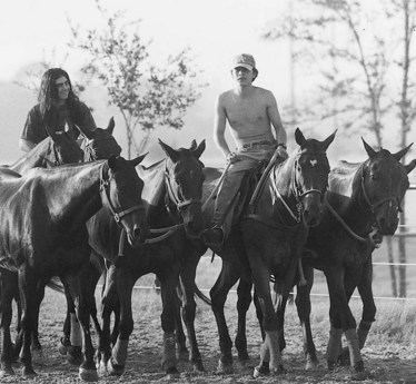
Neck reining is paramount in training a polo prospect because the rider uses only one hand for control. Wide range of movement, the ability to stop and turn quickly, and the ability to exhibit rapid bursts of speed are required. How well and smoothly the horse performs these maneuvers often determines the number of years the horse stays sound and competes successfully. In addition to schooling, fitness training consists of daily galloping (legging up). Ponies often are tied together in sets of four to five. This time-saving practice teaches the horses to travel more calmly together in close contact but can result in traumatic injuries to the lower limbs (Figure 119-2).


Polo ponies are shod with special rim shoes in front that allow for traction and pivoting without applying excessive torque to the lower limb. Medial and lateral heel calks on the hind shoes are helpful for stopping abruptly but often result in coronary band and pastern region lacerations to other horses during competition. For safety reasons, the Great Britain Polo Association only permits a lateral calk on each hind shoe, whereas the United States Polo Association allows medial and lateral heel calks. The size limit for calks is regulated but seldom enforced. Therapeutic corrective shoeing is problematic in playing horses (those actively being used in polo competition) because they may lose traction and maneuverability. Some common shoeing modifications include squared, rolled, and rocker toes; elevated and full-shod heels; and padded soles. The standard support and protection afforded the horse during exercise are leg wraps and coronet boots. All legs are wrapped with double-layer rolled cotton bandages. Impact-resistant European racing boots may be added to cover the metacarpal regions to protect against mallet and hoof trauma. More recently, especially on previously injured limbs, cotton leg bandages have been replaced with neoprene fabric wraps that extend below the fetlock joint and provide additional support. Despite these additional protective barriers, horses may still injure tendons and receive skin lacerations during a game or practice.


Drug testing of polo ponies is not yet compulsory in the United States, and no mention of prohibitive medication is addressed in the United States Polo Association rulebook; however, limited testing is done in Great Britain and France. Attending veterinarians often work for many competing teams within the same tournament, and prompt assessment of injuries is important. Low doses of nonsteroidal antiinflammatory drugs (NSAIDs) are commonly used, especially in horses with wounds and solar bruising. The general aim is to have as many sound horses as possible sharing the workload during a match to avoid the practice of double-chukkering (using the same horse for two chukkers).


Minor conformation abnormalities in polo ponies often can be overlooked, but some faults predispose ponies to specific injuries. Long toes and underrun heels may result in tendonitis of the deep digital flexor tendon (DDFT) and palmar foot pain. Toed-in horses are prone to develop lateral suspensory branch desmitis, whereas toed-out horses are more likely to injure the medial branch. Horses with long pasterns and long third metacarpal bones (McIII) are at increased risk of tendonitis of the superficial digital flexor tendon (SDFT).


The most common sources of lameness in polo ponies are similar to those seen in most other equine sports. Polo ponies are at higher risk for traumatic injury because of the high-impact play and the practice of tethering of horses in close proximity to other horses during shipping, exercise, and polo tournaments. Causes of lameness often seen concurrently include palmar foot pain and PSD and osteoarthritis (OA) of the fetlock joint with chronic suspensory branch desmitis.


Lameness Examination


Horses should be stabled overnight so that they cannot warm out of subtle lameness. The horse is first examined in the stall and then as it walks from the stall. The horse is observed at a trot in a straight line on a hard surface and is circled in both directions. Most polo ponies are reluctant to lunge. If necessary, the horse may be observed under saddle, but Argentine ponies generally resist trotting when ridden.


A systematic examination at rest is begun with the hoof and hoof tester evaluation and then continued proximally in the limb, noting evidence of pain, swelling, or obvious injury. Findings always should be compared with the contralateral limb, especially when palpating the body of the suspensory ligament (SL). Joints are assessed for range of motion and a painful response to flexion. Lower limb flexion is followed by carpal or upper hindlimb flexion. Walking the horse briefly between flexion tests to allow an aggravated response to wear off is wise.


One of us (PJM) attends to many older polo ponies that have effusion of the metacarpophalangeal joints, manifest a positive response to flexion, and even may have visible and radiological evidence of OA, but lameness often is abolished using low palmar digital analgesia. Palpation may reveal one or more fractured splint bones with callus, but the rest of the limb should be examined because the cause of lameness may be elsewhere.


If a definitive diagnosis cannot be made, diagnostic analgesia is performed. Because drug tests are not performed, local anesthetic solutions can be used for diagnostic purposes in actively competing horses. When performing a nerve block, it is important to remember that the block may affect a larger area than intended, primarily related to diffusion of local anesthetic solution to surrounding tissue. High palmar analgesia can mask middle carpal joint pain, and palmar nerve blocks performed at the base of the proximal sesamoid bones (PSBs)can eliminate pain associated with the fetlock joint. For this reason, the horse should be observed at the trot shortly after injection of local anesthetic solution and then again after an appropriate wait.


To reduce time and money spent on lameness diagnosis, one author (PW) prefers to block large areas during the initial examination. Specific blocks then are performed, if necessary, the following day. For example, a horse that shows neither a sensitivity to hoof tester examination nor an increased digital pulse amplitude may go sound after palmar nerve blocks performed at the base of the PSBs. The following day the same horse may show slight improvement after palmar digital analgesia and a 100% improvement with intraarticular analgesia of the distal interphalangeal (DIP) joint. However, two authors (PJM and MWR) prefer to start distally and work proximally in a systematic fashion.


Intraarticular analgesia is used extensively in polo ponies because it is more specific than perineural analgesia. Although intraarticular analgesia requires aseptic preparation and carries a small risk of infection, clients are generally receptive. If lameness is localized to a specific joint on clinical examination, therapeutic agents such as corticosteroids and hyaluronan can be added to local anesthetic solution to confirm diagnosis and initiate treatment simultaneously. One author (PW) uses combination diagnostic and therapeutic arthrocentesis typically in the DIP and proximal interphalangeal (PIP) joints. The horse’s immediate response to local analgesia is noted, and response to therapy is usually evident 2 to 3 days later. Another author (PJM) frequently uses combination diagnostic and therapeutic injections in the DIP and distal hock joints. Combination injections can also be used simultaneously to diagnose and treat back pain. The dorsal aspect of the dorsal spinous process and the interspinous space can be infiltrated with a combination of local anesthetic solution, Sarapin (High Chemical Company, Levittown, Pennsylvania, United States), and a corticosteroid. Response to infiltration is evaluated immediately by riding the horse after injection, and response to medication is evaluated over the next several days.


Because metacarpophalangeal joint disease and splint bone injury are common sources of pain in the polo pony, one author (PJM) prefers specifically to differentiate these sources of pain by first performing intraarticular analgesia of the metacarpophalangeal joint and then later performing a low palmar block. If low palmar analgesia is performed first, both potential sources of pain are eliminated. If pain is detected on palpation of bony exostoses of the splint bones, these areas can be blocked first, before a systematic blocking strategy is followed. One author (PJM) refers to this as the splint block. This block is performed by first blocking the palmar metacarpal nerve distal to the exostoses. If improvement is not seen, the palmar metacarpal nerve just proximal to the exostoses is then blocked (2 mL of local anesthetic solution). A biaxial splint block can be performed if exostoses are found medially and laterally. This block should be done well below the origin of the SL to clearly differentiate PSD from splint bone disease. Splint disease, mainly from direct trauma from mallets and calk trauma, is common in hindlimbs. Diagnostic analgesia is performed as described in the forelimb.


Hindlimb PSD has become a common diagnosis because we are now more aware of it. In the United Kingdom, a variation of the high plantar nerve block, analgesia of the deep branch of the lateral plantar nerve (see Chapter 10), is commonly used to diagnose PSD. Three milliliters of local anesthetic solution is injected deep to the proximal aspect of the lateral splint bone, and 2 mL each is placed over the medial and lateral plantar nerves. If this block is unsuccessful in abolishing pain, each hock joint compartment is blocked subsequently. This procedure then is followed by fibular and tibial nerve blocks. In Argentina, chemical neurolysis (long-term nerve block) of the fibular and tibial nerves frequently is performed for horses with distal hock joint pain or PSD (PJM).


Undiagnosed Lameness


In some horses the lameness is inconsistent and/or subtle, and diagnostic analgesia cannot be performed. Nerve trauma on the abaxial aspect of a PSB may cause episodic, transient severe lameness. An option in a horse with mild inconsistent lameness is to treat with phenylbutazone (2 g orally [PO] twice daily) for 5 days and then reassess the horse. If lameness resolves and does not return after treatment is discontinued, the horse gradually is put back into work. In horses with inconsistent lameness that fail to respond to rest or therapy, we recommend nuclear scintigraphic examination. Exercise intensity can be increased in horses with subtle lameness but is done so with caution. Lameness may become more apparent to the point at which diagnostic analgesia can then be performed. In a horse with recurrent episodes of hindlimb lameness, the veterinarian should be aware of the possibility of an ilial stress fracture (SK).


Occasionally, a polo pony becomes acutely non–weight bearing, with lameness lasting only a few minutes and resolving before examination is possible. If this sort of episode becomes recurrent in the same limb and physical examination reveals no clinically significant findings, we refer the pony for nuclear scintigraphic examination.


Consultation with colleagues and second opinions are always options. It is also important to consider the option of extended turnout. Because the career of a polo pony can last 12 to 15 years, owners are often willing to give the horse 6 to 12 months of turnout to avoid any further injury. If subtle lameness resolves with phenylbutazone therapy, the polo pony can compete because there is no drug testing in polo competition. However, this option must be elected with caution.


Several lameness problems may exist simultaneously in a polo pony, a fact that makes observing the primary or baseline lameness difficult. Subtle signs such as the failure of a horse to stop appropriately, a horse that jumps on after stopping, or a horse that turns one way or the other when stopping (which is probably from outside hindlimb pain; horses turn away from lameness) may reflect low-grade lameness. If these observations have been made, having the horse ridden to witness the problems first hand is useful. In one author’s experience (PJM), the most common source of pain in this type of situation is from the distal hock joints.


Imaging Considerations


Conventional, computed, and digital radiography are the mainstays of imaging, with the front feet and front fetlock joints and hock joints being examined most frequently. The introduction of computed and digital radiography has provided great advantages because, with the exception of faulty positioning or movement, obtaining nondiagnostic radiographs is almost impossible. Exposure can be adjusted at the time of processing, and subtle details that would have been difficult to see on conventional radiographs can now readily be detected and scrutinized easily with computed and digital radiography. Images can be enlarged, and the contrast and brightness can be improved, which are important factors in the diagnosis of incomplete fractures.


Scintigraphic examination is particularly useful in polo ponies with undiagnosed lameness and in those with palmar foot pain, but it is not always helpful in horses with chronic lameness (SK). Motion-correction software has been an important innovation.


Ultrasonography is extremely important in evaluating the damage and healing processes in tendons and the SL in forelimbs and hindlimbs. Transverse images are used more frequently in identifying lesions, whereas longitudinal images aid in assessing healing. Ultrasonographic evaluation of the supraspinous ligament is often useful in horses with obscure hindlimb lameness. We have not found thermography particularly useful in our practices.


Magnetic resonance imaging (MRI) has become available in university settings and in some private equine practices. Despite a long learning curve in the interpretation of magnetic resonance images, many differential diagnoses and prognoses have emerged. Nowhere is this more evident than in obscure foot and pastern lameness with no detectable radiological abnormalities.


Diagnostic arthroscopy in horses with OA of the metacarpophalangeal or carpal joints can be valuable in evaluating the condition of joint surfaces. Palmar intercarpal ligament injury has been diagnosed in ponies with lameness localized to the middle carpal joint but lacking radiological and scintigraphic abnormalities. Tenoscopy and bursoscopy also can be useful diagnostically and therapeutically.


Superficial Digital Flexor Tendonitis


SDF tendonitis is the most common soft tissue injury seen in polo ponies and is by far the most common reason for early retirement. SDF tendonitis can be divided into three categories by location on the limb—high (proximal), midmetacarpal, and low (distal)—or by cause: trauma, speed, and fatigue.


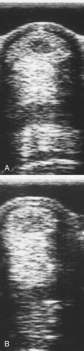
We believe that most peripheral injuries of the SDFT result from tendon trauma while the limb is bearing weight. However, the Editors note that peripheral injuries commonly are seen in other sports horses, such as Standardbred racehorses and reining horses, in which direct trauma is usually not a factor. These injuries occur much more frequently at the midmetacarpal region on the lateral aspect and to a lesser extent on the palmar surface of the tendon (Figure 119-3). Proximal SDF tendonitis also can be caused by trauma (PJM). These areas have a high degree of exposure to swinging mallets and flying hooves. Despite new protective boots, the SDFT is still traumatized with surprising frequency. Traumatic tendon injuries are generally noticed 1 to 2 days after the incident and are characterized by a slight widening of the tendon (not a banana-shaped profile). Lameness is usually not present, but the area is warm and tender to palpation. Some horses have recurrent heat and swelling that resolves quickly with topical and systemic antiinflammatory therapy. Peripheral tendon fiber lesions may involve 20% or less of the cross-sectional area (CSA) of the tendon. However, careful ultrasonographic examination of the medial and lateral borders of the SDFT and critical evaluation of longitudinal images are necessary. Recurrent tendonitis leads to typical swelling and later lameness commonly found with moderate or severe SDF tendonitis.


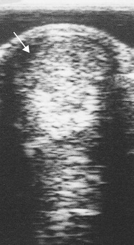
Core lesions and lesions of the SDFT adjacent to the DDFT are thought to be injuries related to speed and fatigue. Hard, fast ground may be a predisposing factor. SDF tendonitis may result in a banana-shaped profile of the metacarpal region. Core lesions compromising between 20% and 25% of the CSA of the SDFT are serious, and the risk of recurrence is high (Figure 119-4). Horses with small CSA tears that extend more than 2.5 cm in length or those with distally located tendonitis involving SDFT impingement by the palmar annular ligament (PAL) are at high risk of recurrence. Despite appropriate therapy, these horses often have chronic and recurrent lameness, and ultrasonographic evaluation reveals a lesion that often fails to heal.


Initial therapy for any polo pony with tendonitis regardless of location or cause includes the application of ice boots and cold-water hosing, sweats, and compression wraps, as well as the administration of NSAIDs. A combination of injection and rest or surgical management has been successful (MWR).


Medical Management


Peritendonous injections over lesions and intralesional injections have become the mainstay of today’s preferred therapy for SDF tendonitis. The aim of this therapy is to return the injured segment of tendon to its elastic state and maintain tensile strength. Peritendonous injections of short-acting corticosteroids and hyaluronan may help to reduce inflammation and improve the cosmetic appearance of the tendon profile, but it is important that corticosteroids are not injected directly into a tendon. Peritendonous corticosteroid injection is frowned on in the United Kingdom (PJM). Autologous adult mesenchymal stem cells, platelet-rich plasma (PRP), and porcine bladder matrix (PBM) have emerged as the biological-based therapies of choice and have markedly increased the success of medical management. One author (PW) prefers an ultrasonographic guided injection of PRP directly into lesions and prefers to use only small volumes of the prepared serum into the lesion. Results using adult fat-derived stem cells are equally satisfactory, but this technique requires preinjection surgical collection, as well as transit time, and is substantially more expensive than using in-house processed PRP. PBM is commercially available (ACellVet, ACell Inc., Columbia, Maryland, United States), requires no surgical harvesting or transit time, and is comparable in price to PRP processing, although it occasionally causes postinjection flares. These flares can be reduced by using a small volume of PBM rather than using the entire reconstituted vial. Daily topical icing and rebandaging, along with NSAIDs, are good precautionary therapies to reduce reactions.


Ultrasonographic assessment of tendons 2 months after injection with one of these biologically based products has revealed remarkable filling within hypoechogenic lesions on transverse images. At 6 months after treatment there is good fiber alignment seen on longitudinal images. The real endorsement of success is that approximately 80% of horses with first-time bowed tendons treated with these products have returned to the same level of polo without recurrence of injury within 8 to 12 months.


Jun 4, 2016 | Posted by in EQUINE MEDICINE | Comments Off on Lameness in the Polo Pony

Full access? Get Clinical Tree

Get Clinical Tree app for offline access