Lameness in Foals

Chapter 128Lameness in Foals


Like all horses, foals are prone to injury and lameness; however, considering the immature and fragile musculoskeletal system, the incidence of serious injury is surprisingly low. Exposure of a foal’s naive immune system to the array of pathogens places the foal at high risk of infectious causes of lameness. Degree of lameness in foals ranges from subtle and nearly imperceptible to non–weight bearing. Although no published studies document the incidence of lameness in foals, the economic impact on the horse industry is substantial when factors such as cost of diagnosis and treatment, long-term morbidity and mortality, and the effect on athletic potential are considered. Economic and emotional losses may be substantial for horse owners and breeders, enough so that they may abandon the horse industry. Most lameness is self-limiting, however, and in many instances early recognition and prompt treatment makes a difference in costly medical bills and improves the long-term soundness or survival. It is imperative that veterinarians learn to recognize the early clinical signs of these serious disorders and to initiate prompt treatment. Clients should be made aware of the potential danger of a wait-and-see approach and a delay in treatment.


Evaluation of a Lame Foal


Foals often resist handling and limb manipulation, making assessing responses difficult and potentially confusing. It is imperative that the veterinarian is patient when evaluating foals. The basics of lameness localization are the same as in adult horses. The stance and any obvious swelling or sensitive areas on palpation should be noted. It is important to palpate the contralateral limb to gauge the foal’s response to manipulation of that limb. Hoof and limb temperature (hot or cold) and digital pulse amplitude should be evaluated. Detailed assessment with hoof testers is invaluable in differentiating causes of hoof lameness. Any aberration in hoof development (e.g., club foot, deformed foot, unbalanced feet, or mismatched sizes of feet) should be noted. Postural or conformational changes, including sudden angular limb deviation, limb laxity, or contraction, may result from altered load bearing associated with lamemess or from structural changes from injury. Careful palpation of all structures of the skeletal system, including all palpable surfaces of joints, bones, digital flexor tendons, and ligaments, with the foal in a standing position and with each limb flexed is important. Swelling should be differentiated as intraarticular, periarticular, or both.


Gait deficits in foals manifest like those in adult horses in that lameness is expressed as reduced time or weight on the lame limb.1 The phase of the stride in which the gait is altered is the result of reducing loading of the limb during the painful portion of locomotion. Foals are more likely to show lameness in dramatic fashion by carrying a limb (i.e., not bearing weight) or exaggerating efforts to reduce load on the limb. It is important to observe the foal at a walk to discern subtle differences in stride and foot placement that may be missed at a trot.


Routine ancillary diagnostic tests used in foal lameness evaluation include diagnostic analgesia; radiographic, ultrasonographic, and nuclear scintigraphic examinations; and arthrocentesis and aspiration of other fluid pockets.1-4 Diagnostic analgesia should be a routine component of lameness localization in foals just as in mature horses, unless a fracture is suspected. Any areas of suspect pathological bone conditions should be evaluated radiologically. Weekly follow-up radiological evaluation may be required to recognize bony changes. Soft tissue evaluation is aided by ultrasonography, especially when the veterinarian is looking for fluid pockets or alterations in soft tissue architecture. Nuclear scintigraphy has limited applications for skeletal evaluation in foals because normal radiopharmaceutical uptake in the physes can confuse or mask subtle abnormalities.


Noninfectious Causes of Lameness


Noninfectious causes of lameness include external and internal traumatic injuries and developmental or metabolic musculoskeletal diseases. Lameness also may result as a sequela to vascular disorders or may be immune-mediated secondary to an infectious problem.


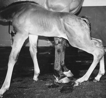
Lameness resulting from external trauma varies with the area involved and the severity of the injury. Essentially any portion of the skeletal system is prone to injury. Traumatic injuries resulting in lameness are common but fortunately self-limiting in most instances. Boisterous activity, coupled with a hazardous environment, places a foal at an increased risk of sustaining injury compared with an adult horse. Trauma from other horses (a mare kicking a foal or collision injuries with other foals) is a common cause of lameness. Impact with stationary objects while a foal is being chased may result in serious injury. Internal trauma includes overexertion injuries, such as a foal running excessively with a mare or being chased, resulting in fracture of the proximal sesamoid bones (PSBs), or rupture of the suspensory apparatus or other soft tissue structures (Figure 128-1). Common fractures in foals include those involving the long bones, physes, PSBs, carpal and tarsal cuboidal bones, and the distal phalanx.1,2,5,6


Fractures of Long Bones


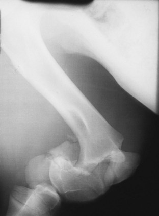
Clinical evaluation may be diagnostic in foals with lameness of traumatic origin. Unstable fractures or severe soft tissue injuries cause obvious, immediate clinical signs. If a fracture is suspected, radiographs are necessary to confirm the diagnosis and determine the severity of the injury (Figure 128-2).


Fractures of long bones most often result from external trauma such as a kick or having the limb pinned beneath an object while rolling. A limb misloaded while rearing may result in fracture of the tibia, femur, or pelvis. If a limb is unstable distal to the femur or humerus because of a fracture, the limb should be immobilized before transport. Surgical approaches and repair techniques for individual fracture types are described elsewhere.


Prognosis for a foal after fracture depends on anatomical location of the fracture; the complexity, orientation, and degree of soft tissue injury around the fracture; and whether the fracture is open. Secondary problems associated with prolonged lameness or confinement and immobilization often dictate the degree of long-term soundness. These problems include excessive laxity of the metacarpophalangeal or metatarsophalangeal joint, varus deviation, or deformity of the foot, such as an underslung or crushed heel and overgrowth of the toe. Mechanical laminitis is not as common as in adult horses, but it does occur. Contracture or excessive laxity of the fractured limb associated with disuse may prevent future soundness.


Fractures of the radius, ulna, and tibia are generally more amenable to surgical repair than are femoral and humeral fractures. If economics do not allow surgery, conservative management of foals with femoral and humeral fractures by stall confinement is a feasible alternative to surgical intervention. Teaching the foal to support its weight on a straw bale or butt bar may alleviate load on the contralateral limb for hindlimb fractures. Complications encountered after surgical repair of femoral or humeral fractures, such as implant failure or infection, are usually fatal. Without postoperative complications, however, a foal has a greater chance of soundness for light athletic use with surgical management. Fractures of the radius and tibia require surgical stabilization if any displacement of the fracture is present. Most foals have a fair to good prognosis for light to medium athletic use or for breeding purposes. Foals with fractures of the ulna that have any displacement should undergo surgery, but those with nondisplaced fissure fractures do not require surgical fixation. Foals with nondisplaced fractures of the ulna should have stall confinement for a minimum of 8 to 10 weeks and undergo radiological monitoring every other day for the first 2 weeks and then weekly to ensure displacement does not occur. Foals with displaced, unstable fractures of the metacarpal and metatarsal bones generally have a guarded to poor prognosis for future athletic use, with or without surgery. Foals with simple, stable fractures of the metacarpal and metatarsal bones not requiring surgery have a good prognosis.


Fractures of the Pelvis


Pelvic fractures in foals are common and usually results from a foal misloading a hindlimb when rearing or from flipping over and landing on one side. Gait and degree of lameness depend on the portion of the pelvis involved and the amount of trauma to the surrounding tissue. Common sites of injury include the tuber coxae, tuber ischium, acetabulum, and the shaft of the ilium. Deep palpation of the area may elicit pain.


The characteristics of the lameness may reflect the site of injury. A foal with a fracture of a tuber ischium is reluctant to advance the ipsilateral hindlimb and has a shortened cranial phase and prolonged caudal phase of the stride. As with many pelvic injuries, the tail often is elevated and held to the opposite side, and the foal may carry the limb when running but bear full weight while walking. Conversely, foals with fractures of a tuber coxae have a shortened caudal phase of the stride, and the injured tuber coxae is often lower than the contralateral side.


Treatment for foals with pelvic fractures includes confinement for 8 to 12 weeks followed by a gradual return to controlled exercise. Improvement in the degree of lameness should be seen within 2 to 3 weeks, depending on the region of the pelvis injured. Foals with acetabular injuries may require a longer duration of confinement before improvement is seen. If the foal is fully weight bearing, controlled walking for short periods after 3 weeks can be started if the foal is tractable.


Prognosis for foals with pelvic fracture not involving the acetabulum is fair to good for light use but guarded for competitive athletics. Some horses are able to race following healing of pelvic fractures. Most foals with pelvic fractures involving the acetabulum have a poor prognosis for an athletic future; however, they generally survive to make breeding animals. In the Editors’ experience, the prognosis for athletic function is not invariably poor because, in contrast to adult horses, foals have a remarkable capacity for bone remodeling, and even some severe articular fractures can heal satisfactorily, allowing normal athletic function.


Fractures of the Physes


Physeal fractures have been categorized and defined using the Salter-Harris classification scheme.2-7 A type I fracture occurs through the physis and involves only the zone of hypertrophied chondrocytes; the adjacent epiphysis and the metaphysis are not involved. A type II fracture occurs across the physis and extends into a portion of the metaphysis. A type III fracture involves the physis and epiphysis and extends into the joint. A type IV fracture includes involvement of the physis, epiphysis, and metaphysis. A compression injury to the physis is referred to as a type V fracture. This classification scheme is applicable to pressure physes that contribute to the longitudinal growth of bone and are subject to compressive forces. Traction physes, such as the olecranon growth plate, are subject to tensile forces and are not included in this classification scheme.


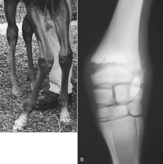
Physeal fractures are common because physeal bone is weak compared with diaphyseal bone. Diagnosis of physeal injuries is made on clinical and radiological findings (Figure 128-3). Clinical signs include varying degree of lameness, pain on palpation of the injured area, swelling, and possibly instability of the limb or angular deviation of the limb distal to the fracture. The primary differential diagnosis for a stable physeal fracture is infectious physitis.


Surgical repair is necessary in foals with displaced, unstable physeal fractures. The major difficulty in surgical stabilization is the short section of epiphysis available for purchase. This shortcoming generally is overcome with the use of a form of T-plate, cross pins, transfixation pins, or tension-band wiring. If adequate reduction and stability are achieved, the prognosis for future soundness is good.


Jun 4, 2016 | Posted by in EQUINE MEDICINE | Comments Off on Lameness in Foals

Full access? Get Clinical Tree

Get Clinical Tree app for offline access