Tim G. Eastman
Keratomas
Keratomas are benign tumors of the hoof that generally arise between the hoof wall and third phalanx as a result of abnormal proliferation of keratin-containing tissue and squamous epithelial cells. Frequently preceded by a subsolar abscess or trauma, these masses arise most often along the coronary band at the toe or the quarters of the equine digit. As the size of the keratoma increases, lameness becomes evident because of pressure exerted on the sensitive laminae and surrounding structures. By the time horses are presented for evaluation, lameness is typically graded at 3 to 4 out of 5.
Diagnosis
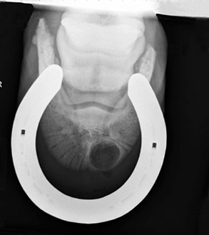
Horses are generally presented for progressive lameness in the affected limb, often with a visible deviation of the coronary band or abnormal hoof growth overlying the region containing the keratoma. No breed or sex predilection has been observed, although forelimbs appear to be more commonly affected than hind limbs. The degree of sensitivity to hoof testers can vary, depending on the location of the mass. If a bulge is visible at the coronary band, that area is often painful to palpation. By use of perineural anesthesia, the lameness is localized to the foot, frequently at the level identified by a basilar sesamoid nerve block. Radiography often reveals a semicircular to circular well-demarcated area of radiolucency along the solar margin of the third phalanx caused by pressure necrosis from the keratoma (Figure 197-1).
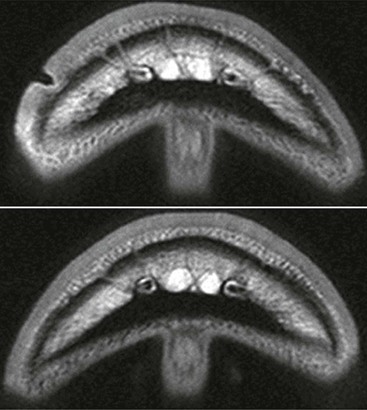
However, because lesions are not always appreciable radiographically, ultrasound, nuclear scintigraphy, computed tomography (CT), and magnetic resonance imaging (MRI) have also been advocated as alternative imaging modalities (Figure 197-2). Advanced imaging with CT or MRI may also be useful in guiding the surgical approach to a keratoma, allowing for a smaller hoof wall resection. Although diagnosis is frequently made presumptively on the basis of clinical signs, histologic evaluation may be performed to confirm the diagnosis and to rule out other types of neoplastic or nonneoplastic conditions.
< div class='tao-gold-member'>
Stay updated, free articles. Join our Telegram channel

Full access? Get Clinical Tree


