Introduction

CHAPTER 1 Introduction


ACCURATE DIAGNOSIS


The major objective of this book is to teach enough of the morphologic and physiologic features of the nervous system to enable the student to make an accurate localization of the lesion in the nervous system. This is the anatomic diagnosis. The differential diagnosis is totally dependent on the anatomic diagnosis and that, in turn, determines the ancillary procedures that will be prioritized to arrive at the most accurate presumptive clinical diagnosis and the subsequent selection of treatment.


The diagnosis of clinical neurologic disorders starts with recognition of the problem—the clinical signs exhibited by the patient and your neurologic examination. This visual and hands-on experience is difficult to learn by reading text descriptions. Direct contact with the affected patient is the ideal teaching model but is impractical in a teaching environment. The most effective alternative is to visualize the clinical signs using video technology. This third edition includes linkage to a website consisting of 381 videos that provide the student with the classical appearance of the common disorders of the nervous system of domestic animals. The anatomic diagnosis is determined by the nature of the problem; that is, clinical signs that you have observed. You should first attempt to determine whether all the clinical signs can be explained by a lesion at one site in the nervous system, a focal lesion, because they are more common than multifocal or diffuse disorders. Based on this anatomic diagnosis, you will next establish a list of disorders that must be able to affect the anatomic location of the lesion. This is the differential diagnosis. You will learn various ways to remind yourself of the disorders to consider. One way is the MIIND system (malformation, injury, inflammation, neoplasia, and degeneration).


Degenerations


Degenerations include the deterioration of cells due to lack of blood supply (ischemia), abnormal cellular metabolism caused by an inherited cellular defect, exposure to exogenous toxins, and abnormalities in other body systems (renal disorders with uremia, diffuse liver disorders with hyperammonemia, cardiorespiratory disorders with hypoxia). Abiotrophy is cell degeneration due to an intrinsic defect in the essential metabolism necessary for the survival and function of that cell, the neuron.


Do not forget to consider the breed of your patient and the possible inherited disorders that must be considered. You will prioritize these disorders in your differential on the basis of signalment, history, and course of the clinical signs and the characteristics of the various disorders being considered.


Based on this ranking of the differential diagnosis, the most useful ancillary procedures will be selected to further confirm or deny the diagnosis under consideration. This selection is especially critical now that neuroimaging by computed tomography and magnetic resonance are available to veterinarians. These procedures require general anesthesia and the costs to the owner of the patient are considerable. Therefore, it is crucial that the correct anatomic diagnosis be made prior to the selection of the ancillary procedures. Your knowledge of the characteristics of the disorders of the nervous system will then permit you to offer a therapy where it is appropriate and a prognosis.


NEURON


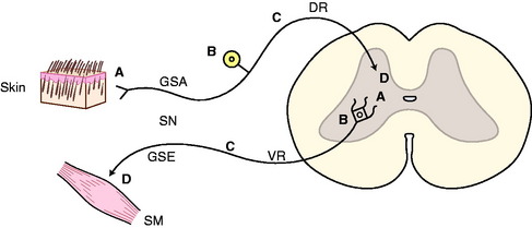
The nervous system is composed of primary functional cells—the neurons and supporting cells, which include the glia and ependyma. In this book the neuron is defined as consisting of a dendritic zone, axon, cell body, and telodendron. The dendritic zone is the receptor portion, where a stimulus from the internal or external environment is converted into an impulse in the neuron. The axon is the cell process composed of neurofilaments that course from the dendritic zone to the telodendron. The telodendron is the termination of the neuron where the impulse leaves the neuron. It is often referred to as the synapse. This synapse may lie at an effector organ or at another neuron. The cell body consists of the nucleus and the major organelles necessary for the neuron to function and may be located anywhere along the axon.


For example, a sensory neuron in the peripheral nervous system for general proprioception may have its dendritic zone in a neuromuscular spindle in a skeletal muscle where it is stimulated by a stretching of the muscle. The axon courses toward the spinal cord through a specific peripheral nerve, then through the dorsal or ventral branch of one of the spinal nerves and into its dorsal root. It then enters the spinal cord and passes into the dorsal gray column of that spinal cord segment to synapse on a second neuron in a nucleus within that gray column. The telodendron is the nerve ending at the synapse on another neuron in that nucleus. The neuronal cell body is located in the spinal ganglion associated with the dorsal root that the axon coursed through to reach the spinal cord. It is actually intercalated in the axon at this point (Fig. 1-1).


Stay updated, free articles. Join our Telegram channel

Aug 26, 2016 | Posted by in INTERNAL MEDICINE | Comments Off on Introduction

Full access? Get Clinical Tree

Get Clinical Tree app for offline access