Infectious Diseases of the Gastrointestinal Tract

Chapter 6 Infectious Diseases of the Gastrointestinal Tract


CALVES


Escherichia coli


Escherichia coli is a normal inhabitant of the gastrointestinal tract of warm-blooded animals and ubiquitous in the farm environment. Disease caused by E. coli in calves may present as enteric or septicemic illness and is an important cause of neonatal mortality in dairy calves. Failure of passive transfer and management practices that allow exposure of the neonatal calf to large numbers of E. coli are of central importance in the pathogenesis of disease. A plethora of E. coli serotypes exist on dairy farms. These gram-negative organisms are classified based on various serologic and antigenic parameters, including cell wall or somatic (O) antigens, capsular (K) antigens, pilar or fimbrial (F) antigens, and flagellar (H) antigens. Heretofore, pilus antigens were sometimes classified as K antigens, but recent reference to pilus antigens as F antigens reduces confusion in this area.


Septicemia (Septicemic Colibacillosis, Colisepticemia)


Etiology.


Colisepticemia in neonatal calves can be considered a disease of poor management. Failure of passive transfer is the primary risk factor for this disease. Colostral transfer of immunoglobulins may be compromised by short dry periods, preparturient leaking of colostrum, assumption that a calf has nursed colostrum just because it is left with the dam for 24 hours, primiparous heifers that have poor-quality colostrum, and many other factors. In addition, poor maternity area and poor calf pen hygiene promote exposure of the calf to the multitude of strains of E. coli capable of causing septicemia. Filthy conditions, calving areas that are dirty, wet, overcrowded, or overused, and failure to dip navels are additional factors that predispose to this problem. Sanitation and hygiene with respect to collecting, storing, and administering colostrum are also emerging as important factors in the provision of adequate passive transfer and the prevention of colibacillosis.


Invasive E. coli of many subgroups are capable of opportunistic, septicemic infection of neonatal calves. Various reviews suggest an involvement of a multitude of possible E. coli types. Variations may be explained by geographic or environmental differences.


Calves with less than 500 mg IgG/dl are very prone to septicemic E. coli, and those with 500 to 1000 mg IgG/dl are defined as having partial failure of passive transfer and are also at increased risk. Adequate passive immunoglobulin that ensures at least 1000 IgG mg/dl serum (10 mg/ml serum) or preferably 1600 mg/dl serum is likely to prevent the disease.


Septicemia caused by E. coli most commonly occurs from 1 to 14 days of age. The onset of disease tends to occur earlier in this timewhen frame calves are exposed to high numbers of E. coli soon after birth (i.e., in the maternity pen). Poor or nonexistent transfer of passive immunoglobulins to the calf also hastens the onset of disease. Invasive E. coli may gain entrance through the navel, intestine, or nasal and oropharyngeal mucous membranes. Once invasion and septicemia occur, clinical signs develop rapidly and usually are apparent within 24 hours. Calves with partial failure of passive transfer or those exposed to less virulent E. coli strains may develop more chronic signs of disease over several days.


Septicemic calves shed the causative E. coli in urine, oral secretions, nasal secretions, and later in the feces, provided they survive long enough to develop diarrhea. Thus transmission may occur among communally housed calves, crowded calves, or uncleaned maternity stalls because of the heavily infected secretions of sick and septicemic calves. Because septicemic calves can shed large numbers of the organism before clinical signs are evident, contamination of communal pens and common-use feeding devices (e.g., esophageal feeding tubes) and direct contact with the infected calf or its feces or urine may promote spread of infection. Infected calves allowed to remain in the maternity area will amplify the level of environmental contamination, thereby placing other neonates born in that area at risk. Similar amplification may occur in calf housing areas and reinforces the biosecurity need for spatial and temporal separation between occupants, as well as the appropriate and routine disinfection of calf housing.


Clinical Signs.


Peracute signs of depression, weakness, tachycardia, and dehydration predominate when highly virulent strains of E. coli cause septicemia. Affected calves usually are less than 7 days of age and may be less than 24 hours old. Although often present early on, fever is usually absent by the time obvious clinical signs of disease occur, when endotoxemia and the resultant poor peripheral perfusion often render the animal normothermic or hypothermic. Exceptions to this rule are calves with peracute disease that collapse when exposed to direct sunlight on hot days—such calves can be markedly hyperthermic. Signs of dehydration are mild to moderate in most cases. The suckle reflex is greatly reduced or absent, and the vasculature of the sclerae is markedly injected. Petechial hemorrhages may be visible on mucous membranes and extremities, particularly the pinnae of the ears (Figure 6-1). The limbs, mouth, and ears are cool to the touch. Affected calves show progressive weakness and lethargy, often becoming comatose before death. Diarrhea is often seen but may not be apparent in peracute cases.
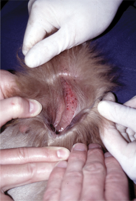


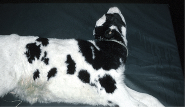
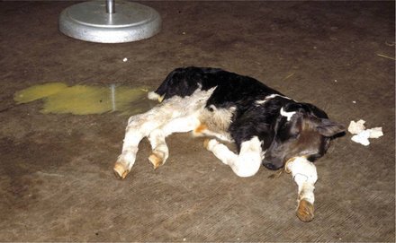
Evidence of localization of infection in certain tissues may become apparent in cases that survive the acute disease. Hypopyon may be present, as may uveitis, which is evidenced by miotic pupils with increased opacity to the aqueous fluid (“aqueous flare”). Hyperesthesia, paddling, and opisthotonus (Figure 6-2) are signs suggestive of septic meningitis. Lameness may result from bacterial seeding of joints and/or growth plates. Signs of omphalophlebitis may be present. Weakness, poor body condition, and recumbency secondary to weakness or joint or bone pain may be present in chronic cases.


Clinical signs of acute septicemia may be difficult to differentiate from those of acute enterotoxigenic E. coli (ETEC) infection because dehydration, weakness, and collapse may be common to both. However, septicemic calves tend to be less dehydrated and have less watery diarrhea than calves with ETEC diarrhea; further, diarrhea tends to develop in the terminal stages of septicemia. Historical data may indicate other neonatal calves have recently shown similar signs or died at less than 2 weeks of age. Other differential diagnoses for acute colisepticemia include asphyxia or trauma during birth, simple hypothermia and/or hypoglycemia, septicemia caused by Salmonella spp., and congenital defects of the central nervous or cardiovascular systems. Polyarthritis caused by Mycoplasma spp. is an important differential diagnosis for septic arthritis secondary to colisepticemia but tends to be seen in older calves, and feeding unpasteurized milk is a significant risk factor for Mycoplasma disease. Salt poisoning, hypoglycemia, congenital neurologic disorders, traumatic injuries, and intoxications (e.g., lead) should be considered as differential diagnoses for meningitis secondary to colisepticemia. We have also seen several herds in recent years with young calves presenting with neurologic signs indistinguishable from meningitis for which the ultimate diagnosis was ionophore toxicity caused by overdosing before feeding milk replacer. Failure of passive transfer and meningitis were not involved.


Ancillary Data.


Calves suffering from peracute E. coli septicemia often have elevated packed cell volumes resulting from dehydration and endotoxic shock. The total white blood cell (WBC) count is variable but is frequently low or within normal ranges. Generally a left shift is observed, and toxic changes (e.g., azurophilic cytoplasm, nuclear hypersegmentation, and Dohle bodies) are often apparent on cytologic examination of blood neutrophils. Plasma fibrinogen concentration is variable. Hypoglycemia is a common finding, and metabolic acidosis, although common, usually is less severe than in calves recumbent as a result of ETEC. In fact, an acid-base and electrolyte determination that does not demonstrate a severe metabolic acidosis in a recumbent, diarrheic, dehydrated calf less than 14 days of age is a grave sign and usually portends septicemia. Severe hypoglycemia would be the only other laboratory finding that might indicate an easily treatable condition. Blood cultures provide the greatest specific diagnostic aid, but results may not be forthcoming in time to help the patient.


Acute, subacute, and chronic septicemic calves may have detectable clinical signs of localization of infection that allow a more definitive diagnostic test (e.g., cerebrospinal fluid [CSF] tap for patients showing signs of meningitis or joint tap to confirm septic arthritis) (Figure 6-3). In chronic cases, the serum immunoglobulin concentration (and serum total globulin concentration) may be normal or increased as a result of de novo synthesis of antibodies in response to the well-established bacterial infection.


Diagnosis.


Whenever clinical signs suggest the diagnosis, the calf’s serum immunoglobulin levels should be analyzed. Although adequate levels of IgG do not rule out the disease, calves with IgG .1600 mg/dl serum based on a single radial immunodiffusion test are unlikely to suffer septicemic E. coli infections. Specific laboratory evaluation of immunoglobulin levels is preferable to field techniques when confirmation of failure of passive transfer (FPT) is essential but may not provide rapid results in the field. Therefore even though dehydration may falsely elevate blood protein levels, these may be useful field tests. Adequate immunoglobulin levels are suggested by serum total protein .5.5 g/dl in clinically ill calves. Serum gamma glutamyl transferase (GGT) activity .50 IU/L is a reliable indicator of adequate passive transfer in ill calves, as is development of visible turbidity in the 18% solution of sodium sulfite turbidity test. Several commercial “quick tests” (e.g., Midlands Bio-Products, Boone, IA) are now available for determining IgG concentration in calves.


Blood cultures provide definitive diagnosis but usually provide this information too late to be of practical value. When multiple calves are affected, however, blood cultures can help to differentiate E. coli septicemia from septicemia caused by other pathogens (e.g., Salmonella spp.); this differentiation is relevant for determining the source of infection and initiation of preventive measures. Further, antimicrobial sensitivity testing of blood culture isolates may aid in directing therapy for subsequent cases. Clinicians and producers should be aware of the differences between specific antigenic strains of E. coli (e.g., ETEC) capable of producing severe disease in calves with adequate passive transfer and the everyday, commensal, and environmental E. coli often associated with sepsis caused by FPT. This is an important distinction, lest clients concentrate preventive efforts and management on specific vaccination programs rather than colostrum and neonatal calf management.


Treatment.


Treatment of peracute E. coli septicemia usually is unsuccessful because of overwhelming bacteremia and endotoxemia in the patient. Signs progress so quickly that most septicemic calves are recumbent and comatose by the time of initial examination. Shock, lactic acidosis, hypoglycemia, and multiple organ failure are common in peracute cases.


If treatment is attempted, correction of endotoxic shock and acid-base and electrolyte abnormalities, effective antimicrobial therapy, and nutritional support are the primary goals. Intravenous (IV) balanced electrolyte solutions should contain dextrose (2.5% to 10%) and sodium bicarbonate (20 to 50 mEq/L if the plasma bicarbonate concentration is, 10 mEq/L) to address hypoglycemia and metabolic acidosis. Adjustments of the concentration of dextrose and sodium bicarbonate in polyionic fluids can be guided by subsequent serum chemistry results. Maintaining normoglycemia in some peracute and acute septicemic calves can be extremely challenging due to consumption of administered glucose by bacteria. Antimicrobials used to treat neonatal septicemia should be bactericidal and possess a good gram-negative spectrum, such as ceftiofur, trimethoprim-sulfa, or ampicillin. Parenteral administration is necessary to achieve effective blood concentrations. Aminoglycosides such as gentamicin or amikacin can be used alone or in conjunction with the synergistically acting beta-lactam antibiotics (e.g., ceftiofur, penicillin, or ampicillin). The use of potentially nephrotoxic aminoglycosides in a dehydrated patient with prerenal azotemia must be weighed against the potential bactericidal activity of the drugs. Given the present concerns regarding aminoglycoside use in food animals, use should be limited to situations in which other antibiotics have proven ineffective. Further, a minimum 18-month slaughter withdrawal must be enforced for calves that receive aminoglycosides. Use of fluoroquinolones (e.g., enrofloxacin, danofloxacin) in dairy calves is currently not permitted under federal law in the United States.


If the previous therapy stabilizes the patient, a transfusion of 2 L of whole blood from (preferably) a bovine leukemia virus (BLV) and bovine virus diarrhea virus persistently infected (BVDV-PI) free cow should be performed because failure of passive transfer is assumed or confirmed. This translates to a dosage of 40 ml of whole blood/kg for the calf. Bovine plasma, which is commercially available, may also be used at the same dosage rate as whole blood. Nutritional support ideally entails frequent feedings of small volumes of whole milk or good-quality milk replacer. Partial or total parenteral nutrition (TPN) may be considered for valuable calves, particularly those with concurrent and significant enteritis. Deep, dry bedding, good ventilation, and good nursing care are essential adjuncts to medical treatment.


Specific sites of localized infection also may require specific therapy. As an example, patients manifesting seizures because of meningitis may require diazepam to control seizures. Calves with septic joints often require joint lavage. In many cases, arthrotomy is necessary to remove fibrin clots from infected joints.


Chronic cases usually are cachectic, have polyarthritis and diarrhea, and have an extremely poor prognosis. Although recumbent, weak, dehydrated, and emaciated, these patients tend to have relatively normal acid-base and electrolyte values, so fluid therapy is of limited value.


Prevention: Colostrum and Management.


Sporadic cases of E. coli septicemia are unfortunate events, but endemic neonatal calf losses resulting from this disease demand a thorough evaluation of management regarding dry cows, periparturient cows, and newborn calves. There are two basic questions that require answers: (1) are newborn calves being fed sufficient volumes of high-quality colostrum soon enough after birth? And, (2) is the environment likely to harbor large numbers of E. coli during the periparturient and neonatal period? In other words, two facets of the dairy operation must be carefully critiqued: colostrum management and the hygiene of the maternity area and neonatal calf pens. A few very basic concepts regarding colostrum should be understood:


Given this current summation of colostral quality research, the following recommendations are made for newborn dairy calves:


In addition to colostrum, maternity (calving) pen management practices that predispose to E. coli septicemia must be corrected. The importance of maternity pen hygiene cannot be overstated because no level of passive immunoglobulin transfer can protect completely against gross filth in the environment, and conversely even calves with partial or complete FPT may survive when cleanliness is exceptional. Dry cows should not be kept in filthy environments that allow heavy fecal contamination of the coat and udder. Maternity stalls or calving areas should be cleaned, disinfected, and adequately bedded between uses by different cows.


Newborn calves should be removed from the calving area as soon as possible after birth because they will inevitably incur fecal-oral inoculation as they attempt to nurse. Ideally calves should be moved from the maternity area into individual hutches, without being allowed to contact one another. This may not be feasible on larger dairies with limited manpower. In such situations, a small “safe pen” for calves can be constructed adjacent to the maternity pens. A safe area is a sheltered, fenced-in, well-drained, concrete-floor pen located in or near the maternity area. These typically measure approximately 20 3 20 ft. Walls should be constructed to prevent contact with cows or bedding from the maternity area. This small area can be cleaned and disinfected daily with relative ease, and fresh bedding can be added easily. A large gate to facilitate cleaning with a bucket loader should be installed at one end of the safe pen to facilitate efficient (and therefore regular) removal of all bedding before cleaning and disinfection, which should be rigorous and regularly scheduled. This pen becomes the holding area for all newborn calves in the maternity area. Personnel on the dairy are made responsible for moving newborn calves into the safe pen as soon as possible after birth; use of gloves and footbaths will aid in preventing contaminating newborns with pathogens carried on boots or clothing. The calves are less likely to become rapidly inoculated with maternity area pathogens. The calves are kept here until the calf attendant can provide colostrum and move the calves to hutches. It is critical the safe pen be disinfected regularly and not be used as a long-term housing area for calves, or accumulation of pathogens is inevitable. On large dairies, particularly those experiencing high calf morbidity and mortality problems in the first 2 weeks of life, it may be cost-effective to dedicate one employee to the maternity pen whose sole responsibility is the prompt removal of newborn calves and colostrum administration. Only larger dairies will be able to implement this because 24-hour coverage will be necessary to monitor and care for all calvings.


Enterotoxigenic Escherichia coli


Etiology


ETEC produces enterotoxins that cause secretory diarrhea in the host intestine. Several types of enterotoxins have been identified, and a single ETEC may be capable of producing one or more enterotoxins. Both heat-labile (LT I, LT II) and heat-stable (STa, STb) enterotoxins have been identified in ETEC. In calves, ETEC producing the low molecular weight STa cause the majority of neonatal diarrhea problems.


Pathogenic ETEC must be able to attach to the host enterocytes to create disease. Once adhered, the organism releases enterotoxin, which induces the intestinal epithelial cell to secrete a fluid rich in chloride ions. Water and sodium, potassium, and bicarbonate ions follow chloride, creating a massive efflux of electrolyte-rich fluid into the intestinal lumen. Although some of this fluid is reabsorbed in the colon, the efflux of secreted fluid exceeds the colonic capacity for fluid absorption, and watery diarrhea results.


Because enterotoxins are nonimmunogenic, efforts to control ETEC in calves have centered on inducing antibody against fimbrial proteins. The type I fimbriae that allow pathogenic ETEC to attach to enterocytes are proteins that initially were categorized with capsular (K) antigens. Currently fimbriae are classified as F antigens. Unfortunately even the current literature still refers to K-99, K-88, and so forth, rather than the current designation, F-5 and F-4, respectively. In calves, F-5 (K-99) is the most commonly identified antigenic type and has received the most attention regarding diagnostics and vaccines for calves. However, ETEC possessing other fimbrial antigens or multiple fimbrial antigens including F-41, F-6, and some types still not widely identified are capable of causing diarrhea in calves. Some ETEC possess more than one type of fimbriae, and both F-41 and F-5 types may be isolated from an ill calf. Colostrum possessing passive antibodies against a specific ETEC fimbriae type will protect the newborn calf against that specific F type but will not cross-protect it against others (Table 6-1).


TABLE 6-1 Fimbriae Antigens


Designations
New Old Toxin
F 4 K 88 LT I
F 5 K 99 STa
F 6 987 P STa
F 41   SIa

As for E. coli septicemia, ensuring prompt feeding of adequate levels of colostrum is extremely important to protect calves against ETEC. However, because of lack of cross-protection against various fimbrial antigens, even calves with excellent passive transfer are at risk to ETEC with F types other than those against which the dam has provided colostral antibodies. Colostrum containing antibodies against specific F types will prevent attachment of homologous ETEC to calf enterocytes by coating the fimbriae binding sites. Therefore colostral protection is a local effect of IgG in the gut. To be effective, colostrum containing antibodies against ETEC F antigens must be fed as early in life as possible, lest ETEC colonize the gut before colostrum has been consumed. Although one experimental design showed colostral F-5 antibodies to be effective up to 3 hours after experimental oral challenge with ETEC F-5, it is more practical to assume colostrum should “beat” the ETEC to the gut. Other management factors in addition to colostral feeding are also important in the pathogenesis of ETEC diarrhea. Conditions that allow or encourage buildup of ETEC in the dry cows, in the maternity or neonatal calf facilities, and/or in stored colostrum increase the risk of ETEC diarrhea, as is true with E. coli septicemia. Marrow products may also decrease the risk of ETEC by binding to toxin receptors and/or preventing proliferation of pathogenic bacteria.


Affected calves are usually 1 to 7 days of age, with most cases seen in calves less than 4 to 5 days of age. Calves are most susceptible to F-5 ETEC during the first 48 hours of life and thereafter begin to build resistance to those organisms. Concurrent infection with rotavirus may extend the age of susceptibility to ETEC diarrhea to approximately 10 days of age. In older calves, continued exposure to heavy inocula of pathogenic ETEC may result in intestinal colonization and shedding of the organism in normal or diarrheic stools, thereby facilitating new infections in neonates.


Clinical Signs


These signs vary from mild diarrhea with resultant spontaneous recovery to peracute syndromes characterized by diarrhea and dehydration that progress to shock and death within 4 to 12 hours.


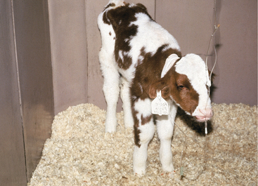
Because of the multitude of ETEC types and variability in their pathogenicity, as well as the influence of passive transfer, individual farms may have sporadic or endemic problems resulting from ETEC. Mild disease is common on many farms and seldom is brought to a veterinarian’s attention. These calves have loose or watery feces but continue to nurse (Figure 6-4). Spontaneous recovery or apparent response to the farmer’s favorite “calf-scour” treatment (usually an oral antibiotic) is the rule. Owners usually call for veterinary assistance only when peracute cases develop, a high morbidity is apparent, calves fail to respond to over-the-counter medications, or mortality in neonatal calves is experienced.


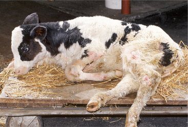
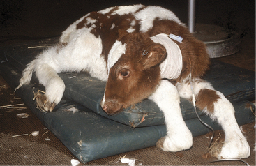
Peracute cases may produce dehydrated, weak, and comatose calves within hours of the onset of the disease. Historically these calves usually have nursed normally and appeared healthy until signs develop. Dehydration and weakness are the predominant signs (Figure 6-5). Mucous membranes are dry, cool, and sticky. The suckle reflex is weak or absent.


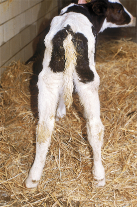
Most peracute cases show evidence of voluminous diarrhea (Figure 6-6), with watery feces coating the tail, perineum, and hind legs. Some calves with peracute disease may not have diarrhea; however, the pooling of fluid in the intestinal lumen creates abdominal distention, and fluid splashing sounds can be detected by simultaneous auscultation and ballottement of the right lower abdominal quadrant. Mild, transient colic may be noted early in the disease course. Bradycardia and cardiac arrhythmia accompany the systemic signs in some peracute cases and result from hypoglycemia or hyperkalemia. Atrial standstill has been documented in some bradycardiac calves with hyperkalemia. Rectal temperatures usually are normal or subnormal if the calf is recumbent.


Because of the profound and peracute signs, differentiation of E. coli septicemia from peracute ETEC infection often is difficult in the field setting. In prodromal, peracute ETEC cases, the presence of massive fluid in the intestine, as evidenced by abdominal contour, simultaneous auscultation and ballottement, and/or abdominal ultrasonography, are key indicators that the characteristic voluminous diarrhea is impending. Further, on resuscitation with IV fluids, ETEC cases typically break with voluminous diarrhea, and provided the concurrent abnormalities in hydration, electrolyte, acid-base, and glucose status are addressed properly with IV fluids, calves with ETEC typically show rapid clinical improvement. In contrast, calves with E. coli septicemia show less voluminous diarrhea, and diarrhea typically develops late in the disease course. Also, unlike ETEC cases, calves with E. coli septicemia typically fail to demonstrate a dramatic clinical response to fluid resuscitation.


Acute cases show obvious watery diarrhea, progressive dehydration, and weakness over 12 to 48 hours. The character and color of the feces vary as well, but feces usually are voluminous, watery, and yellow, white, or green. Such calves may have low-grade fever or normal temperatures and deterioration in the systemic state and suckle response. Continued secretory diarrhea gradually worsens the hydration and electrolyte deficiencies; weight loss is apparent—especially if fluid intake is decreased by reduced suckling.


Translocation of bacteria from the gut into the systemic circulation is an uncommon event when ETEC is the sole agent involved because these organisms do not invade the deeper layers of the gut wall and incite minimal intestinal inflammation. Therefore evidence of localized infection (e.g., hypopyon, arthritis) is uncharacteristic of ETEC infection and more indicative of colisepticemia and/or septicemia secondary to other enteric diseases.


Clinical Pathology


Peracute infections resulting from ETEC cause severe secretory diarrhea that result in a classical metabolic acidosis with low plasma bicarbonate and low venous pH. Hyperkalemia and hypoglycemia also are characteristic. Hyperkalemia results from efflux of K1 from the intracellular fluid in exchange for excessive H1 in the extracellular fluid (ECF). Reduced renal perfusion contributes to retention of K1 in the ECF. Mild hyponatremia and hypochloremia are inconsistently present. Dehydration is generally greater than 8%, and corresponding elevations in packed-cell volume (PCV) and total protein are typical. The WBC count usually is normal, although elevated numbers of WBC may be present because of extreme hemoconcentration, and stress leukograms occasionally are discovered. Leukopenia, left shifts, and toxic cytologic changes in neutrophils are uncommon in ETEC infections, and those findings more likely support septicemia and/or salmonellosis.


Hypoglycemia is more likely to be present if the interval between feedings is prolonged; this finding is not present in all peracute cases. Blood values for a typical case are shown in Table 6-2. Mild azotemia resulting from prerenal causes (reduced renal perfusion) is common and should be kept in mind when use of potentially nephrotoxic drugs is considered in these patients.


TABLE 6-2 Laboratory Data From a Typical Peracute ETEC Infection in a 7-Day-Old Holstein Calf


Item Tested Electrolyte (mEq/L) Normal Range
Na = 127 132-150
K+ = 8.1 3.9-5.5
Cl = 104 97-106
HCO3 = 12 20-30
Tot CO2 = 10 26-38
Ven pH = 7.09 7.35-7.50


Diagnosis


The diagnosis is suggested by the calf’s age, physical signs, and laboratory data. Peracute ETEC may be difficult to differentiate from E. coli septicemia and salmonellosis in neonatal calves based on clinical signs alone. Response to appropriate fluid therapy strongly supports ETEC infection, as does confirmation of adequate patient serum immunoglobulins.


Definitive diagnosis requires isolation of an E. coli possessing pathogenic F antigens that allow intestinal attachment in calves having typical clinical signs. When submitting samples for culture, the clinician should indicate that ETEC infection is a possibility and should request typing of E. coli isolates for F antigens (by immunofluorescence, slide agglutination, or polymerase chain reaction [PCR]) and, if available, for enterotoxin (by PCR or, rarely, by ligated gut loop assays). In fatal cases, ETEC can be cultured from the ileum; a section of ileum should be tied off, placed in a sterile container, and transported on ice packs to the laboratory. Isolation of ETEC from diarrheic feces of older calves is generally considered to reflect the presence of the pathogen in the calf population. In such cases, fresh specimens of jejunum and ileum should be examined carefully for histologic evidence of attachment of ETEC to enterocytes. These findings suggest participation in enteric disease by ETEC, rather than simple intestinal colonization by the organism. Obtaining samples for culture before antibiotic therapy, particularly when oral antibiotics are being given, is an important factor in the diagnostic workup of a potential ETEC outbreak.


Histologic examination of fresh samples of ileum and jejunum of affected calves greatly aids in confirming the diagnosis of ETEC infection. Sections of ileum should be cut into 2- to 3-cm lengths, then split longitudinally and swirled in 10% neutral buffered formalin solution to aid in rapid fixation of the mucosa. Samples for histology should not be tied off because this delays fixation of the mucosa. In classic ETEC infection, a dense population of gram-negative rods are found adherent to the mucosa of the ileum.


Because mixed infections with combinations of ETEC, rotavirus, coronavirus, and Cryptosporidium are common, feces and/or intestinal contents should also be analyzed for viral and protozoan pathogens. Salmonellosis also must be included in the differential diagnosis because many types of Salmonella sp. can cause severe diarrhea, dehydration, shock, and acid-base disturbances similar to ETEC. Fever, neutropenia, and a left shift are more commonly observed in Salmonella patients. In addition, enterotoxemia resulting from Clostridium perfringens must be considered, especially in peracute cases with abdominal distention but no diarrhea. Calves with clostridial enterotoxemia may be weak, dehydrated, or “shocky” but seldom have as dramatic a metabolic acidosis as that found in ETEC infections.


Treatment


Appropriate replacement and maintenance fluids constitute the primary therapy of ETEC infection in neonatal calves. Correction of metabolic acidosis and hypoglycemia and reestablishment of normal hydration status are imperative. Calves with peracute signs or those that are recumbent require IV therapy. Calves that can stand but show obvious dehydration, cool and dry mucous membranes, and have a reduced or absent suckle reflex also should initially be given IV therapy. Calves that are ambulatory and have a good suckle response usually can be treated with oral fluids.


Concentrations of required electrolytes based on subjective clinical parameters rather than objective laboratory tests are empiric at best, but sometimes are necessary in field situations. Therefore rules of thumb include:


These rules of thumb are not absolute, and chronic low-grade bicarbonate loss and/or increased D-lactate production in the gut may create profound acidosis over a period of days in a calf having only minimal signs of dehydration. A 40-kg calf that is judged 10% dehydrated will need 4 L of fluid simply to address current needs. For all calculations of replacement electrolytes, a 50% ECF will be assumed for neonates. Therefore a 40-kg calf will be assumed to have 40 3 0.5 5 20 L ECF compartment. If this 40-kg calf has a venous plasma bicarbonate concentration of 10 mEq/L, and 25 to 30 mEq/L is the desired normal level, then 15 to 20 mEq of bicarbonate must be replaced in each liter of ECF. Therefore 20 L 3 15 mEq 5 300 mEq (20 L 3 20 mEq 5 400 mEq) would be necessary to correct the bicarbonate deficit associated with the metabolic acidosis.


Total CO2 of venous blood also may be used to calculate base deficits in lieu of HCO3 values.


Much research data and individual clinical opinions exist as to the most appropriate content of initial fluid therapy for ETEC infections in calves. An effective solution, first proposed by Dr. R. H. Whitlock, is formulated by adding 150 mEq of NaHCO3 to 1 L of 5% glucose. This combination is used for the initial 1 to 3 L of IV therapy, depending on severity of measured or suspected metabolic acidosis. Glucose corrects hypoglycemia if present, and both bicarbonate and glucose facilitate potassium transport back into cells, thereby lessening the potential cardiotoxicity associated with hyperkalemia. Some reports minimize the importance of hyperkalemia and suggest using IV potassium in the initial fluid. These workers and others emphasize that dehydrated calves having severe ETEC-induced secretory diarrhea have a total body K1 deficit despite having an elevated ECF [K1]. Although this latter medical fact may be true, it seems risky to tempt fate by administering K1-containing solutions as the initial therapy for a patient known to be hyperkalemic. This is especially true for a patient with bradycardia or arrhythmias because deaths occasionally have occurred when potassium-containing fluids have been given as initial therapy. Once plasma K1 and HCO32 levels are quickly improved by the initial 1 to 3 L of 5% dextrose with 150 mEq NaHCO3/L, potassium-containing fluids can be safely used. Balanced electrolyte solutions such as lactated Ringer’s solution suffice for maintenance fluid needs, but supplemental NaHCO3 and dextrose may be required to address continued secretory losses and anorexia. Response to treatment usually is dramatic in calves with ETEC secretory diarrhea. Calves initially recumbent usually appear much improved following 2 to 4 L of appropriate IV fluids and usually can stand within 6 hours and begin to nurse within 6 to 24 hours of initial therapy. This type of prompt response strongly suggests a correct diagnosis and tends to rule out septicemia because septicemic calves seldom respond promptly, if at all. Depending on the setting (field versus clinic), maintenance or intermittent IV fluid therapy may be continued or replaced by oral fluids in those calves that quickly regain a suckle response and are eager to eat.


Antibiotic therapy for peracute ETEC infections remains controversial, with current concerns focused on antimicrobial residues in edible tissues and indiscriminate and unnecessary use of antimicrobials leading to resistance. However, in peracute cases, the overlap of many clinical signs with colisepticemia often prompts the clinician to include antimicrobial treatment in the therapeutic regimen. In a Canadian study, diarrheic calves, 5 days of age were found to be at significantly greater risk of bacteremia than older calves; this age range obviously includes calves at risk for ETEC infection. Further, in cases with fever and severe debilitation, the veterinarian is often prompted to consider the possibility of complicating conditions such as bacterial pneumonia. Oral antimicrobial treatment offers the potential benefit of reducing the number of ETEC in the gut, and by reducing the source of enterotoxin, one might reduce the drive for hypersecretion. In his thorough review of the subject, Constable found published evidence supporting the logic and clinical efficacy of amoxicillin trihydrate (10 mg/kg orally every 12 hours) or amoxicillin trihydrate–clavulanate potassium (12.5 mg combined drug/kg orally every 12 hours) for at least 3 days for treatment of undifferentiated calf diarrhea. Repeated use of these products over the long term is likely to induce resistance; therefore long-term efforts must focus on prevention rather than treatment.


Recommended treatments for diarrheic calves with signs of severe systemic illness (e.g., fever, weakness that persists after fluid resuscitation) include ceftiofur (2.2 mg/kg intramuscularly [IM] or subcutaneously [SQ] every 12 hours), amoxicillin, or ampicillin (10 mg/kg IM every 12 hours). Extra-label drug use regulations apply to all of these regimens. Systemic antibiotics usually are continued for 3 to 5 days based on the calf’s clinical response, temperature, and character of the feces. Most ETEC that result in high calf mortality have limited antibiotic susceptibility, and sensitivity testing or MIC levels should be determined when the herd history or clinical data suggest high morbidity and mortality from ETEC.


Feces usually remain more watery than normal for 2 to 4 days. If diarrhea persists beyond this time, concurrent infection with other organisms is likely. Other treatments for peracute cases may include flunixin meglumine (1.1 mg/kg IV or SQ every 24 hours) directed against potential endotoxemia, resolution of fever, and reduction of pain associated with fluid-filled bowel. Repeated dosages of this product carry the risk of renal and gastrointestinal injury because continued use of flunixin meglumine interferes with vasodilatory prostaglandin synthesis in the gut and kidney.


Milk or milk replacer should be withheld for no more than 24 to 36 hours, during which time a high-quality oral electrolyte energy source may be fed several times (four to six times) daily. Holding ETEC-infected calves off milk or replacer for prolonged times creates weight loss from inadequate energy intake and places calves at risk of starvation. Even though many oral electrolytes are supplemented with dextrose as an energy source, no commercial oral electrolyte solution provides enough energy for maintenance needs, especially for dairy calves in hutches during winter weather. Weight will be lost, and starvation may occur if these electrolyte solutions are fed as the only ration for more than 1 or 2 days. In highly valuable calves undergoing hospitalization, treatment with parenteral nutrition offers an excellent option to at least approximate maintenance calorific needs while the calf is nil per os (NPO). Calves with ETEC are so significantly catabolic that they will still lose weight despite calorific supplementation with IV lipid and amino acids. Careful monitoring of blood glucose for hyperglycemia and strict attention to aseptic technique, as well as catheter and fluid line maintenance, are important when administering parenteral nutrition. Consequently it is rarely practical outside of a referral hospital.


The alkalinizing potential of oral electrolyte solutions is of great importance, especially when those solutions are utilized as ongoing therapy for peracute cases following initial IV fluids, or when those solutions are used as sole therapy of less severely affected calves having ETEC. Continued HCO32 loss accompanying ETEC secretory diarrhea must be anticipated and treated. Therefore oral electrolyte solutions containing bicarbonate are most helpful. The optimal oral electrolyte solutions typically possess 70 to 80 mEq of alkalinizing potential per liter (as bicarbonate or acetate), dextrose, and electrolytes; these should be fed at 4 to 6 L/day. Oral electrolyte solutions that when mixed with water are nearly isotonic are preferred over those that are markedly hypertonic.


Concerns regarding adding oral electrolyte solutions to milk or milk replacers revolve around the alkalinizing solutions’ tendency to interfere with abomasal clot formation. Therefore oral electrolytes are fed during separate feedings at least 30 minutes before or after a milk feeding. Calves do not digest sucrose effectively, and addition of table sugar to “home remedy” electrolyte mixtures will reliably worsen fluid and electrolyte losses in diarrheic stools. After 24 to 36 hours of oral electrolyte treatment, calves may be fed small volumes of milk or milk replacer. Calves that respond rapidly to initial fluid resuscitation can be started back on small volumes of milk or milk replacer at an earlier time. During recuperation, calves should be deeply bedded in dry straw or similar bedding material and provided shelter from rain and snow. When milk feedings are resumed, feedings are best performed in small volumes frequently. If this is not possible, total milk or replacer should be divided into two to three daily feedings. Supplemental oral electrolyte solutions can be continued if ongoing fluid and electrolyte losses are assumed to result from continued diarrhea, and these solutions should be fed at intervals between milk or replacer feedings. Unless the calf is hypoglycemic or acidotic, isotonic electrolyte solutions are preferred because they allow a more normal abomasal transit than do hypertonic solutions.


Treatment of acute ETEC infections in calves that are ambulatory and still able to suckle may not require IV therapy. cessation of milk or replacer feeding coupled with substitution of oral electrolyte-glucose solutions for 24 to 36 hours may be sufficient. Bicarbonate loss and resulting metabolic acidosis should not be underestimated, however. It is imperative to use highly alkalinizing electrolyte glucose solutions to provide 4 to 6 L of fluids per day. Parenteral antibiotics are indicated if the affected calf is febrile, and oral antibiotics may be administered when the herd medical history indicates involvement of a highly pathogenic ETEC. Milk or replacer should be restored after 24 to 36 hours, and electrolyte feedings should be used as fluid supplements in the intervals between milk feedings.


Mild ETEC infections seldom require veterinary care. Spontaneous recovery is the rule, and supportive care with oral electrolyte solutions frequently is used by owners in such cases. Use of over-the-counter remedies is widespread among dairy farmers treating mild ETEC infections or nonspecific “calf scours.” Although little scientific evidence is found to justify these products, anecdotal testimonials from farmers exist for oral neomycin, tetracyclines, sulfas, and other antibiotics, oligosaccharides, or protectants. Over-the-counter calf diarrhea products that contain methscopolamine, atropine, or products that reduce intestinal motility are contraindicated and may cause bloat and ileus if overdosed. Bismuth subsalicylate is palatable and can be used safely in calves.


Prevention


This assumes prime importance when a high morbidity, significant mortality, or both occur on a dairy farm. It is not unusual to encounter 70% to 100% morbidity and mortality when virulent strains of ETEC are present. These strains also tend to be resistant to many antibiotics. The usual situation is that the owner tries multiple over-the-counter products on the first few affected calves and then calls for veterinary assistance to select a “better” antibiotic. One or more calves may die or require intensive therapy before a thorough investigation of the problem ensues.


The veterinarian must avoid the temptation to simply provide or suggest a “newer” or better antibiotic if the problem is to be solved. Feces must be submitted from more than one acutely affected calf. If necessary, bull calves should be raised in the identical manner as heifers just to allow them to develop disease and allow early sampling. A qualified diagnostic lab must identify the E. coli as an ETEC stain with attachment antigens and determine antibiotic susceptibility.


Management must be meticulously assessed as to cleanliness of dry cows, colostrum, feeding instruments, maternity areas, and newborn calf facilities. Evidence of successful passive transfer of immunoglobulins must be evaluated in several consecutive calves to rule out E. coli septicemia or poor colostral feeding as the major cause of ETEC infection. Culturing of colostrum at milking and from the bucket or bottle immediately before its feeding can be used to assess the cleanliness of colostrum milking procedures, colostrum storage, and feeding instrument hygiene. Readers are directed to the previous section on colisepticemia for more details on assessment of colostrum management.


If an ETEC with attachment antigens such as F-5 is cultured from the feces of more than one affected calf, preventive measures can be instituted. Management factors including colostral feeding must be emphasized, lest preventive vaccines are looked on by the farmer as a “silver bullet” that obviates any need for management changes. When specific F antigen ETEC are involved, a commercial bacterin containing these F types can be administered to the dry cows 6 weeks and 3 weeks before freshening or at manufacturer’s recommended times. Autogenous bacterin manufacturers should be required to show data on endotoxin levels in bacterins because administration of endotoxin-rich vaccines to adult cattle can cause dramatic production losses and/or abortion. Calves born to dry cows in the next few weeks that are unlikely to form sufficient colostral antibodies in response to bacterins may be given commercially available oral monoclonal antibodies (Genecol 99 (E. coli antibodies), Schering-Plough Animal Health Corp., Union, NJ) against F-5 if this is confirmed as the attachment factor for the ETEC in question. Monoclonal antibody products must be given immediately after birth before colostrum is fed. Valuable calves at risk born to these same dry cows also may receive systemic antibiotics for the first 3 to 5 days of life in an effort to prevent infection with the ETEC identified, and selection of appropriate antibiotics should be based on antibiotic susceptibility testing of the causative organism.


Rarely a particular serotype of E. coli other than the F-5 pilus type is isolated from the small intestine of scouring neonatal calves. If the organism subsequently is consistently confirmed as the pathogen (based on samples from multiple affected calves) and commercial dry cow vaccines have not altered the incidence of disease, an autogenous bacterin should be considered. However, the use of autogenous bacterins can only be justified when an absolute diagnosis of a highly pathogenic ETEC has been confirmed by isolates from several affected calves and commercial bacterins fail to stop the disease. Because free endotoxin content may be high in some autogenous vaccines made from gram-negative organisms, the manufacturer should “wash” the preparation to reduce endotoxin content, and data on endotoxin content in the final product should be requested. It is important to resist the temptation to initiate autogenous bacterin production using a nonspecific E. coli isolate obtained from one or more calves that merely had colibacillosis as a result of FPT.


Other Escherichia coli Diarrhea


Etiology


Although less common than ETEC, other forms of E. coli have been identified as causes of clinical calf diarrhea. Enteropathogenic are defined as those capable of attachment and effacement of intestinal cell microvilli. Attaching and effacing (AEEC) are EPEC that do not produce enterotoxins but may produce cytotoxins of various types. They do not possess Shigella-like invasiveness. These organisms have been isolated from calves with diarrhea that have histologic evidence of effacement of microvilli in the cecum, colon, and distal small intestine. cellular degeneration may ensue if the organisms produce cytotoxins. These histologic changes enable differentiation of AEEC from ETEC that attach to enterocytes but do not cause histologic damage. Because the lesions typically involve the large intestine, dysentery and diarrhea may be observed. Malabsorption, maldigestion, and protein loss are characteristic of disease with AEEC or EPEC. Calves from 2 days of age up to 4 months of age may be infected, and other enteric pathogens often are present concurrently.


Shiga-like toxin-producing E. coli (SLTEC) are another type of E. coli that produce hemorrhagic colitis and the hemolytic uremic syndrome in humans. These organisms also have been called EHEC and occasionally have been found in calves. Some of these strains invade the mucosa to reside in the lamina propria of the large intestine and produce a severe hemorrhagic colitis. Ulcerative colitis with hemorrhage may be present grossly and microscopically in necropsy specimens. Those producing Shiga-like toxin (verotoxin) create enterotoxemia, inhibition of protein synthesis, and vascular damage in the involved intestine. Other less common strains of E. coli produce cytotoxic necrotizing factor (CNF), a potent cytotoxin that may be linked genetically to a plasmid that encodes fimbriae and toxins. This plasmid may be found in the same strains of E. coli responsible for calf diarrhea and septicemia in neonatal farm animals.


Treatment


Therapy is similar to that for ETEC infection except that whole blood transfusions of 2 L of blood may be necessary in calves with severe dysentery and fecal blood loss. ceftiofur is the most frequently used parenterally administered antimicrobial for this disease. Broad-spectrum antibiotics such as gentamicin (6.6 mg/kg SQ or IV every 24 hours), amikacin (15 mg/kg SQ or IV every 24 hours), or trimethoprim-sulfa combinations (22 mg/kg IV or orally every 12 hours) are also used because of the microvillus or mucosal damage to the intestine, but these represent extra-label drug use in the United States. Prognosis is guarded for calves with AEEC or SLTEC infections unless intensive care is provided. Colonic, cecal, or distal ileal pathology may be so severe as to cause ulceration or perforation of the intestine in some cases. Because of the gross and histologic intestinal pathology, corticosteroids and prostaglandin inhibitors are contraindicated except when used once, in conjunction with initial shock therapy, because these drugs reduced cytoprotective mechanisms of the bowel.


Because of the maldigestion and malabsorption created by these organisms, oral electrolyte-energy sources may be less useful than in ETEC. These products, however, usually are recommended for at least the first 36 to 48 hours of therapy. Calves continuing to have diarrhea after 48 hours can be returned to milk or replacer feeding but may be candidates for TPN if they are valuable enough to warrant the expense.


Rotavirus


Etiology


Rotaviruses are members of the Reoviridae family and are classified further via complicated division into groups (serogroups), serotypes, and subgroups. The rotaviruses cause diarrhea in multiple species, including humans. Although the rotaviruses share certain antigens and cross-infection of species occurs with some strains, in general resistance is specific, and cross-protection against heterologous strains is poor.


Calves usually are infected by group A serotypes and less commonly by group B serotypes. Initially identified by Mebus and co-workers, the Nebraska rotavirus isolate was used extensively for study and vaccine production. Other group A serotypes have been identified in the United States and abroad. Exposure to rotaviruses apparently is widespread in the cattle population based on serologic surveys. Older calves and adult cattle serve as carriers of the virus, shedding the virus intermittently in feces. In addition, up to 20% of healthy calves may shed rotavirus. As a rule, rotaviruses coexist with other neonatal enteric pathogens such as ETEC and C. parvum in herd calfhood diarrhea outbreaks. Experimental mixed infections of rotavirus with bovine virus diarrhea virus (BVDV) have been shown to result in more severe diarrhea than infection with either of these agents alone, suggesting some synergistic effect in pathogenicity.


Neonatal calves (,14 days of age) are at greatest risk for infection by enteric rotavirus, and most infections occur during the first week of life. Prevalence of infection in neonatal calves born on dairy farms harboring the virus is high, morbidity is high (50% to 100%), and mortality varies greatly. Clinical manifestations of disease and mortality in calves are influenced by several factors, including level of immunity to the virus, magnitude of viral inoculum, viral serotype, concurrent infection of the gastrointestinal tract or other systems, stress, and crowding. Germ-free calves infected by rotavirus have self-limiting diarrhea and rapid recovery. Infected calves in field situations may have inapparent, mild, moderate, or fatal disease. As is true with most enteric pathogens, the younger the patient, the higher the likelihood of severe disease because of losses of water, electrolytes, and body nutrient reserves secondary to diarrhea.


Rotavirus infection is limited to the small intestine and characterized by destruction of villous enterocytes and subsequent replacement of these columnar cells by immature and more cuboidal cells derived from the intestinal crypts. Although these new immature cells are resistant to further viral infection, they are unable to carry out the normal digestive and absorptive tasks necessary for villous enterocytes because of deficient disaccharidase and sodium-potassium ATPase activities. Therefore rotavirus diarrhea is characterized by maldigestion and malabsorption. To further complicate matters, the intestinal crypt cells continue their normal secretory function, which is no longer balanced by absorptive villous function. Thus net secretion outweighs absorption and contributes further to diarrhea. Increasing intraluminal osmotic pressure also may draw further water into the bowel as lactose and other undigested nutrients pass through the gut and are fermented in the colon to volatile fatty acids. Bacterial fermentation of undigested lactose creates both D- and L-isomers of lactic acid; in diarrheic calves, absorption of the slowly metabolized D-isomer may result in accumulation of this acid in the systemic circulation, thereby contributing to the development of metabolic acidosis. Water and electrolyte losses of variable severity occur in affected calves.


The level of local passive immunity conferred to calves by colostral intake somewhat determines the risk and relative severity of infection. Colostrum with a high virus-neutralizing antibody titer (.1:1024) against rotavirus is protective against experimental infection. However, unless colostrum or colostrum/milk combinations with titers this high continue to be fed, this local protection “wears off” within a few days, and the calf becomes susceptible to infection. Colostrum or colostrum milk/combinations with lower virus neutralizing titers may impart partial protection. Feeding of colostrum having very high levels of IgG1 antibodies against rotavirus soon after birth may establish high circulating humoral antibodies against rotavirus. Although this humoral protection will not, by itself, protect a calf from infection, a portion of these IgG1 antibodies are secreted back into the intestine over time and are thought to confer additional local protection against infection.


Treatment


Treatment is nonspecific and generally follows therapy described for ETEC regarding indications and types. Several differences are noted, however:


2. Maldigestion, as well as malabsorption, will influence the duration of diarrhea and digestibility of milk or milk replacers in viral enteritis patients. Once diarrhea from rotavirus becomes evident, the damage to the intestinal lining has already occurred, and only time and supportive care can allow the intestine to heal. Nutritional support is a critical component of that supportive care—particularly because rotaviral scours may persist for 3 to 7 days. Producers should be counseled that provision of milk or milk replacer is necessary in viral enteritis, even though the maldigestion of the milk nutrients may contribute in part to the pathologic process. Denial (for .24 hours) of milk feeding to a calf with viral diarrhea places the calf at significant risk for cachexia and may lower its resistance to opportunistic disease. Death from starvation may occur in such cases, particularly during times of inclement weather (Figure 6-7). To quote Dr. Chuck Guard, “If a calf scours for a week, and all that the calf is fed is oral electrolyte replacer, then that calf will be well hydrated and will have absolutely perfect blood electrolyte concentrations and acid-base balance on the day it starves to death.” Producers should learn to live with the “more-in, more out” rule: The more milk goes in the front end, the more diarrhea comes out the back end. However, this process is not necessarily harmful because digestion and absorption of some fraction of milk nutrients is likely to occur, and these nutrients are necessary to support the tissue synthesis required to return the intestine to normal. Any exacerbation of fluid losses and acidosis that may result from maldigestion of milk nutrients can be offset by aggressive fluid and electrolyte replacement. Ideally the affected calf should be fed small amounts frequently with the addition of lactaid tablets!


IV fluid therapy is necessary for recumbent, extremely dehydrated, or “shocky” patients, and patients that have lost their suckle reflex. IV fluid therapy is best guided by acid-base and electrolyte determinations. If this is not practical or available, however, the most severely affected calves with acute diarrhea should be assumed to have metabolic acidosis, low bicarbonate, high potassium, and low glucose values. Guidelines for fluid therapy are available in the section on treatment of ETEC. Parenteral nutrition may be “life saving” in calves with cachexia.


Although there is no need for antibiotic therapy in pure rotaviral enteritis, the likelihood of mixed infections and the pathologic damage to enterocytes that fosters attachment of bacterial pathogens may be reason enough to treat severely affected calves with systemic antibiotics.

Stay updated, free articles. Join our Telegram channel

Apr 26, 2017 | Posted by in GENERAL | Comments Off on Infectious Diseases of the Gastrointestinal Tract

Full access? Get Clinical Tree

Get Clinical Tree app for offline access