Infectious Canine Hepatitis and Canine Acidophil Cell Hepatitis


Infectious Canine Hepatitis and Canine Acidophil Cell Hepatitis


Craig E. Greene


Infectious Canine Hepatitis


Etiology


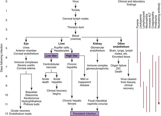
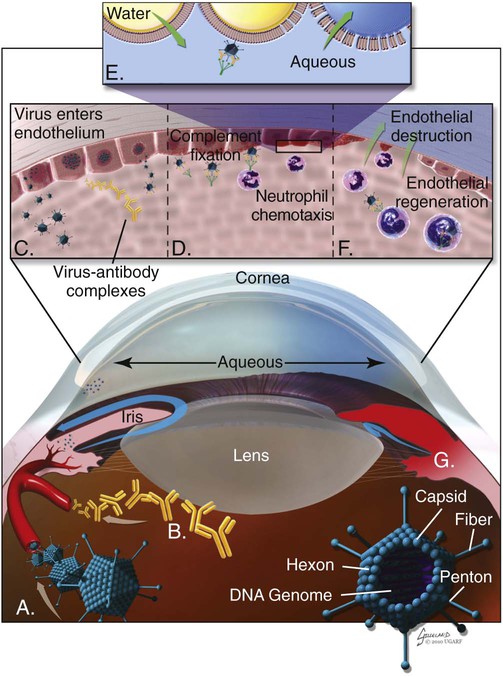
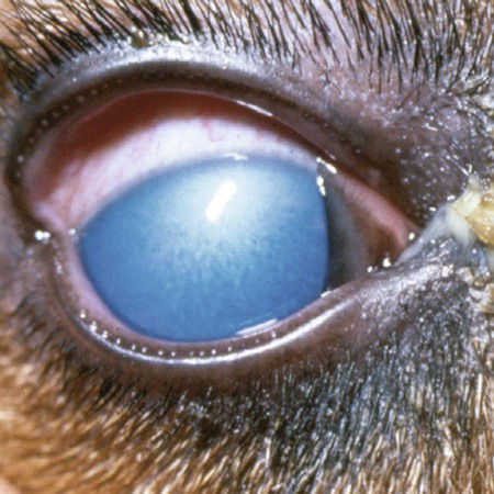
Infectious canine hepatitis (ICH), caused by canine adenovirus (CAV)-1, has worldwide serologic homogeneity, as well as immunologic similarities to human adenoviruses. Historical synonyms for the disease include fox encephalitis and Rubarth’s disease. CAV-1 is antigenically and genetically distinct from CAV-2, which produces respiratory disease in the dog (see Etiology, Chapter 6). Genetic variants of CAV-2 have been isolated from the intestine of a puppy with hemorrhagic diarrhea and from kenneled dogs with diarrhea. Human adenoviruses have been used as vectors for recombinant vaccine testing in dogs.26


As with other adenoviruses, CAV-1 is highly resistant to environmental inactivation, surviving disinfection with various chemicals such as chloroform, ether, acid, and formalin, and is stable when exposed to certain frequencies of ultraviolet radiation. CAV-1 survives for days at room temperature on soiled fomites and remains viable for months at temperatures below 4° C. CAV-1 is inactivated after 5 minutes at 50° C to 60° C, which makes steam cleaning a plausible means of disinfection. Chemical disinfection has also been successful when iodine, phenol, and sodium hydroxide are used; however, all are potentially caustic.


CAV-1 causes clinical disease in dogs, coyotes, foxes, wolves, and other Canidae and in Ursidae (bears). A fatal infection was reported in an otter (Lutra lutra).31 In addition to these carnivores, serum antibodies have also been detected in marine mammals including walruses (Odobenus rosmarus) and sea lions (Eumetopias jubatus).5,32,32 The high prevalence of naturally occurring serum neutralizing antibodies in the unvaccinated feral and wildlife dog population suggests that subclinical infection is widespread.2,14,16,17,38 CAV-1 has been isolated from all body tissues and secretions of dogs during the acute stages of the disease. By 10 to 14 days postinoculation (PI), the virus can be found only in the kidneys and is excreted in the urine for at least 6 to 9 months. Aerosol transmission of the virus via the urine is unlikely, given that susceptible dogs housed 6 inches apart from virus shedders do not become infected. Viral spread can occur by contact with fomites, including feeding utensils and hands. Ectoparasites can harbor CAV-1 and may be involved in the natural transmission of the disease.


Pathogenesis


After natural oronasal exposure, the virus initially localizes in the tonsils (Web Fig. 4-1), where it spreads to regional lymph nodes and lymphatics before reaching the blood through the thoracic duct. Viremia, which lasts 4 to 8 days postinfection, results in rapid dissemination of the virus to other tissues and body secretions, including saliva, urine, and feces. Hepatic parenchymal cells and vascular endothelial cells of many tissues including the central nervous system (CNS) are prime targets of viral localization and injury.


Initial cellular injury of the liver, kidney, and eye is associated with cytotoxic effects of the virus. A sufficient antibody response by day 7 postinfection clears the virus from the blood and liver and restricts the extent of hepatic damage. Widespread centrilobular to panlobular hepatic necrosis is often fatal in experimentally infected dogs, with a persistently low (less than 4) virus neutralization antibody titer. Acute hepatic necrosis can be self-limiting and centrilobularly restricted such that hepatic regeneration occurs in dogs that survive this phase of the disease. Dogs demonstrating a partial neutralizing antibody titer (greater than 16, less than 500) by day 4 or 5 postinfection may develop chronic active hepatitis and hepatic fibrosis. Persistent hepatic inflammation continues, probably as a result of chronic latent hepatic infection with virus. Dogs with sufficient neutralization antibody titers (at least 500) on the day of infection usually show little clinical evidence of disease. Dogs immune to parenteral challenge with CAV-1 are still susceptible to respiratory disease via aerosolized viral particles.


Both virulent and modified live strains of CAV-1 produce renal lesions. Virus detected by positive immunofluorescence and ultrastructural evaluation initially localizes in the glomerular endothelium in the viremic phase of disease and produces initial glomerular injury. An increase in neutralizing antibody at approximately 7 days postinfection is associated with the glomerular deposition of circulating immune complexes (CICs) and transient proteinuria. CAV-1 is not detected in the glomerulus after 14 days postinfection; however, it persists in renal tubular epithelium. Tubular localization of the virus is primarily associated with viruria, and only a transient proteinuria is noted. A mild focal interstitial nephritis is found in recovered dogs; however, unlike the liver disease, evidence that chronic progressive renal disease results from ICH cannot be found.


Clinical complications of ocular localization of virulent CAV-1 occur in approximately 20% of naturally infected dogs and in less than 1% of dogs after subcutaneous-modified live virus (MLV) CAV-1 vaccination. The development of oculr lesions begins during viremia, which develops 4 to 6 days postinfection; the virus enters the aqueous humor from the blood and replicates in corneal endothelial cells.


Severe anterior uveitis and corneal edema develop 7 days postinfection, a period corresponding to an increase in neutralizing antibody titer (Fig. 4-1). CIC deposition with complement fixation results in chemotaxis of inflammatory cells into the anterior chamber and extensive corneal endothelial damage. Disruption of the intact corneal endothelium, which serves to pump fluid from the cornea into the anterior chamber, causes accumulation of edematous fluid within the corneal stroma.


Uveitis and edema are usually self-limiting unless additional complications or massive endothelial destruction occurs. Clearing of corneal edema coincides with endothelial regeneration and restoration of the hydrostatic gradient between the corneal stroma and aqueous humor. Normal recovery of the eye is usually apparent by 21 days postinfection. If the inflammatory changes are severe enough to block the filtration angle, increased intraocular pressure can result in glaucoma and hydrophthalmos.


Complications are often associated with the pathogenesis of ICH. Dogs are more prone to develop bacterial pyelonephritis as a result of renal damage after ICH infection. Disseminated intravascular coagulation (DIC), a frequent complication of ICH, begins in the early viremic phase of the disease and can be triggered by endothelial cell damage, with widespread activation of the clotting mechanism, or by the inability of the diseased liver to remove activated clotting factors. Decreased hepatic synthesis of clotting factors in the face of excessive consumption compounds the bleeding defect.


Although the cause of death in ICH is uncertain, the liver is a primary site of viral injury. Hepatic insufficiency and hepatoencephalopathy can result in a semicomatose state and death. Some dogs die so suddenly that liver damage with resulting hepatic failure does not have time to occur. Death in these dogs can result from damage to the brain, lungs, and other vital parenchymal organs or from the development of DIC.


Clinical Findings


ICH is most frequently seen in dogs younger 1 one year, although unvaccinated dogs of all ages can be affected. Severely affected dogs become moribund and die within a few hours after the onset of clinical signs. Owners frequently believe that their dog was poisoned. Clinical signs in dogs that survive the acute viremic period include vomiting, abdominal pain, and diarrhea with or without evidence of hemorrhage.


Abnormal physical findings in the early phase of infection include increased rectal temperature (39.4° C to 41.1° C [103° F to 106° F]) and accelerated pulse and respiratory rates. Fever may be transient or biphasic early in the course of the disease. Tonsillar enlargement, usually associated with pharyngitis and laryngitis, is common. Coughing and auscultated harsh lower respiratory sounds are manifestations of pneumonia. Cervical lymphadenomegaly is frequently found with subcutaneous edema of the head, neck, and dependent portions of the trunk. Abdominal tenderness and hepatomegaly are usually apparent in the acutely ill dog. A hemorrhagic diathesis that is demonstrated by widespread petechial and ecchymotic hemorrhages, epistaxis, and bleeding from venipuncture sites can occur. Icterus is uncommon in acute ICH, but it is found in some dogs that survive the acute fulminant phase of the disease. Abdominal distention is caused by accumulation of serosanguineous fluid or hemorrhage. CNS signs, including depression, disorientation, seizures, or terminal coma, can develop at any time after infection. In foxes and rare reports in domestic dog pups, CNS signs can be seen in the absence of other systemic manifestations.7


Mildly affected dogs may recover after the first febrile episode. Clinical signs of these uncomplicated cases of ICH frequently last 5 to 7 days before improvement. Persistent signs may be found in dogs with a concurrent viral infection such as canine distemper or in dogs that develop chronic active hepatitis. More severe or additional clinical signs occur in dogs that are co-infected with other pathogens.9,10,10


Corneal edema and anterior uveitis usually occur when clinical recovery begins and may be the only clinical abnormalities seen in dogs with inapparent infection (also see Infectious Canine Hepatitis in Chapter 92). Dogs with corneal edema show blepharospasm, photophobia, and serous ocular discharge. Clouding of the cornea usually begins at the limbus and spreads centrally (Fig. 4-2) (see Fig. 92-17). Ocular pain, present during the early stages of infection, usually subsides when the cornea becomes completely clouded. However, pain may return with the development of glaucoma or corneal ulceration and perforation. In uncomplicated cases, clearing of the cornea begins at the limbus and spreads centrally.


Diagnosis


Early hematologic findings in ICH include leukopenia with lymphopenia and neutropenia. Neutrophilia and lymphocytosis occur later in dogs with uncomplicated clinical recovery. An increased number of dark-staining (activated) lymphocytes and nucleated erythrocytes may be found. Serum protein alterations, detectable only on serum electrophoresis, are a transient increase in α2-globulin by 7 days postinoculation and by a delayed increase in γ-globulin, which peaks 21 days postinoculation.


The degree of increased activities of alanine aminotransferase (ALT), aspartate aminotransferase, and serum alkaline phosphatase depends on the time of sampling and the magnitude of hepatic necrosis. These enzyme increases continue until day 14 postinoculation, after which they decline, although persistent or recurrent elevations may be found in dogs that develop chronic active hepatitis. Moderate to marked bilirubinuria is frequently found owing to the low renal threshold for conjugated bilirubin in the dog; hyperbilirubinemia is uncommon. Of diagnostic importance, the increase in ALT, a measure of hepatocellular necrosis, is often disproportionately higher than the increase in serum bilirubin, despite the diffuse nature of the hepatic injury. This disparity, which is typical for ICH and differentiates it from most other causes of widespread hepatic necrosis, results from the predominant centrilobular nature of the necrosis around the hepatic venules. In contrast, the periportal, peripheral lobular regions around the bile duct are spared. Measures of hepatic function, such as serum ammonia levels or bile acid retention, can be increased during the acute course of ICH or later in dogs that develop chronic hepatic fibrosis. Similarly, hypoglycemia may be found in dogs in the terminal phases of the disease.


Coagulation abnormalities characteristic of DIC are most pronounced during the viremic stages of the disease.30 Thrombocytopenia with or without altered platelet function is usually apparent. One-stage prothrombin time, activated partial prothrombin time (aPTT), and thrombin time are variably prolonged. Early prolongation of the aPTT probably results from factor VIII consumption. Factor VIII activity is decreased, and fibrin or fibrinogen degradation products are increased. Platelet dysfunction and later prolongation of the aPTT probably result from increased fibrinogen degradation products. Prolongation of the prothrombin time is usually less noticeable.


Proteinuria (primarily albuminuria) is a reflection of the renal damage caused by the virus and can usually be detected on random urinalysis because the concentration is greater than 50 mg/dL. The increase in glomerular permeability can result from localization of the virus in initial stages of infection. Alternatively, as the disease progresses, glomeruli become damaged by CICs or as an effect of DIC. Abdominal paracentesis yields a fluid that varies in color from clear yellow to bright red, depending on the amount of blood present. It is usually an exudate with inflammatory cells and with a protein content greater than 5.29 g/dL (specific gravity greater than 1.020).


Bone marrow cytology reflects the dramatic change in leukocytes in the peripheral circulation. Megakaryocytes are absent or decreased during the viremic stage of the disease, and those that are present may have altered morphology.


Cerebrospinal fluid is within reference limits in dogs with neurologic signs caused by hepatoencephalopathy; it is usually abnormal in dogs that develop nonsuppurative encephalitis from localization of the virus within the brain. Protein concentration (greater than 30 mg/dL) increases with mononuclear pleocytosis (greater than 10 cells/µL). The aqueous humor also has increased concentrations of protein and cells associated with anterior uveitis.


Results of laboratory procedures previously discussed are suggestive of ICH and are the primary means of making a diagnosis in clinical practice. Antemortem confirmation, although not essential for appropriate therapy, can be obtained by serologic testing, virus isolation, and immunofluorescent staining for intralesional virus. Serologic tests include virus neutralization, indirect hemagglutination assay, complement fixation, immunodiffusion, and enzyme-linked immunosorbent assay. These tests usually show higher titers after infection with virulent virus in contrast with that in MLV vaccines.


CAV-1 can be isolated because it is highly resistant and readily replicates in cell cultures of several species, including dogs. Typical adenovirus-induced cytopathology includes clustering of host cells and detachment from the monolayers with the formation of intranuclear inclusions. When viremia begins, on day 5 PI, CAV-1 can be cultured from any body tissue or secretion. The virus is isolated in the anterior chamber during the mild phase of uveitis before antibody infiltration and immune complex formation. Culturing the virus from the liver of dogs is often difficult because hepatic arginase inhibits viral nucleic acid replication. The virus has not been isolated from the liver later than 10 days PI, even in dogs with chronic active hepatitis, perhaps because viral latency develops. The kidney is the most persistent site of virus localization, and CAV-1 can be isolated from the urine for at least 6 to 9 months after the initial infection.


Immunofluorescent techniques are used experimentally to confirm the presence of virus within various tissues. This method has helped locate the sites of viral replication, the spread of the virus within the cells, and the presence of viral antigen in inclusion bodies. Immunoperoxidase procedures, applied to formalin-fixed, paraffin-embedded tissues, have detected virus in liver tissues stored for up to 6 years. Polymerase chain reaction (PCR) techniques have been developed to detect CAV-1 in biologic specimens and to distinguish the virus from CAV-2 in clinical specimens.8,20,20

Only gold members can continue reading. Log In or Register to continue

Stay updated, free articles. Join our Telegram channel

Aug 6, 2016 | Posted by in INTERNAL MEDICINE | Comments Off on Infectious Canine Hepatitis and Canine Acidophil Cell Hepatitis

Full access? Get Clinical Tree

Get Clinical Tree app for offline access