Infectious Arthritis and Fungal Infectious Arthritis

Chapter 65Infectious Arthritis and Fungal Infectious Arthritis


image Infectious Arthritis


Alicia L. Bertone


Classic clinical signs of infectious arthritis are heat and swelling and rapid development of non–weight-bearing lameness, often in less than 24 hours. The suspicion of joint infection increases if a predisposing risk factor is evident, such as prematurity, a high sepsis score, or multisystemic disease in a foal1,2 or preceding joint injection in an adult horse. Fracture and nonarticular infection (cellulitis or foot abscess) need to be differentiated from infectious arthritis, because in these diseases, acute, severe lameness also develops.3,4


Causes


Musculoskeletal infection was reported to cause death in 5.2% of 2468 foals.5 Yearly morbidity was 27.4% (677 foals), and morbidity attributed to musculoskeletal infection was 2.1%. Septicemia was the second most common cause of death, and hematogenous spread was the most common cause of infectious arthritis.6 Bacteremia with infectious arthritis in foals decreased survival.7 The risk of infection was highest in the first 30 days postpartum and was lowest in practices that assessed the efficacy of transfer of passive immunity. Isolation of Salmonella species from synovial fluid and systemic disease were associated with an unfavorable prognosis for survival.1


In two retrospective studies of 153 mature horses8,9 the most common causes of synovial infection were traumatic wounds (36.5% for joints and 55% for tendon sheaths), injections (34.1% and 22%), postoperative infection (1.0% for arthroscopy,10 19.8% overall for joints), and idiopathic causes (9.5% and 22%).9 Standardbreds,9 draft breeds,10 and the tarsocrural joint9,10 were overrepresented in adult horses with joint infection, reflecting a greater number of joint injections in these breeds and this joint.


Of 424 bacterial isolates from 233 horses with joint, tendon sheath, or bone infection, 91% were aerobic or facultative anaerobes. The most common organisms were Enterobacteriaceae (28.8%), followed by streptococci (13%) and staphylococci (11.8%).11 In foals, Enterobacteriaceae including Escherichia coli and Staphylococcus species1,12 were more likely to be isolated. Staphylococci, specifically Staphylococcus aureus, are the most common organisms isolated from infections occurring after surgery or injections. Foals or horses with infectious arthritis secondary to penetrating wounds are likely to have multiple bacterial infections. Clostridium species have been isolated from foals13 and were the most common anaerobes isolated, particularly from wounds near the hoof.11 Fungal or mycobacterial organisms are a rare cause of infectious arthritis but can be considered pathogens if identified in pure culture more than once (see later).14,15 Reactive arthritis in foals with septicemia from Rhodococcus equi or subsequent to injection reactions can be confused with infectious arthritis, but lameness usually is not prominent, and synovial fluid nucleated cell counts are often within normal limits.16,17


Examination and Initial Management


Potential infectious arthritis must be considered an emergency and is treated most effectively with early diagnosis. A systematic approach should include hematological examination; measurement of plasma fibrinogen; synovial fluid analysis, including cytological examination, Gram stain, and synovial fluid culture (up to 5 mL of synovial fluid in a broth culture bottle); and radiography.3 In foals, particularly those with abnormal sepsis scores, blood culture should be performed simultaneously. In adult horses, systemic blood examination is less rewarding than in foals, particularly in the early phases of clinical signs. In horses with experimental infectious arthritis, elevations in leukocyte count and total protein and fibrinogen concentrations have been found to take several days to develop and were changed significantly from baseline values for individual horses, but they remained in the normal range for all horses.18 Before arthrocentesis is performed, surgical scrub materials, sterile gloves, needles, syringes, broth culture bottles, ethylenediamine tetraacetic acid (EDTA) or heparin tubes for cytological examinations, smear slides for Gram stain, and a dose of antimicrobial agents to instill directly into the joint after sampling should be available (see the following discussion).


Adult horses should be sedated. Foals can be placed in lateral recumbency with administration of an α2-agonist and synthetic narcotic combination. Although data suggest that aseptic preparation of unclipped hair may be adequate and may significantly decrease bacterial counts on the skin,19 clipping the site for arthrocentesis is strongly recommended. In horses with periarticular wounds, arthrocentesis should be performed well away from the wound to avoid the risk of joint contamination, if the joint is not contaminated already from the wound. To determine if a joint and wound communicate, the clinician should infuse 50 to 200 mL of balanced electrolyte solution into the joint after a synovial fluid sample has been obtained for culture and cytological examination. The clinician should watch closely for leakage of fluid from the wound. This is easier than using blue dye injections or contrast radiography. The joint should be drained and antibiotic instilled into the joint after samples have been obtained and any lavage or injection of fluid completed. Samples should be submitted immediately for evaluation.


If the joint fluid is grossly cloudy, turbid, or flocculent, broad-spectrum antibiotics initially should be given intravenously (IV), and ingress and egress or through-and-through lavage of the joint should be performed until diagnosis of infectious arthritis can be confirmed. Antibiotic should be instilled into the joint after lavage has been completed. If a wound or puncture is the inciting cause, endoscopic evaluation of the synovial cavity is recommended. In one study of 95 contaminated or infected joints, 43% had intraarticular foreign material found at endoscopy.20


Diagnosis


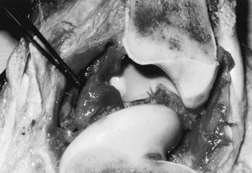
Gross evaluation of the synovial fluid can be informative. If newspaper print cannot be read through the fluid sample, it probably has a cell count of at least 30 × 109 nucleated cells per liter. Fluid from infected joints is usually turbid, cloudy, and watery. Flocculence develops in chronically infected joints or joints that have been invaded with a needle or surgery. Blood contamination makes gross assessment of synovial fluid difficult, and determining if infection is present without clinicopathological analysis is often impossible. Serosanguineous fluid commonly is obtained from infected joints (Figure 65-1). An estimate of the amount of blood (packed cell volume [PCV)] or red blood cell count) can be made to correct for the number of nucleated cells contaminating the sample (hemorrhage from arthrocentesis or leakage of blood from the infection):


image


where WBCs are white blood cells.


Synovial fluid nucleated cell count, differential cell count, and total protein concentration are the most useful parameters to evaluate in diagnosing infectious arthritis. Although other biomarkers, such as lactate, metalloproteinases, and myeloperoxidase activity, have been found to be increased in synovial fluid from infected joints, most correlate with the nucleated cell count.18,21,22 Synovial fluid nucleated cell counts in excess of 30 × 109 cells/L, with more than 80% neutrophils, or total protein concentration in excess of 40 g/L are considered consistent with infection, particularly if these synovial fluid parameters correlate with clinical signs and predisposing circumstances.3,23 The likelihood of isolating the causative organism is correlated positively to the nucleated cell count—that is, higher nucleated cell counts are associated with greater isolation rates.23 Approximately 25% of samples from horses suspected of having infectious arthritis have bacteria on Gram stain, confirming infection and offering the added benefit of assisting with identifying the organism and the initial selection of an antimicrobial agent.18


For horses that do not meet obvious criteria for infectious arthritis, assessment of all parameters is necessary to conclude that the joint is infected. It is important to note that joints can be infected with nucleated cell counts lower than 30 × 109/L. Infection should be suspected if nucleated cell counts are 10 × 109/L to 30 × 109/L and fluid is not serosanguineous, clinical signs of substantial lameness exist, fluid is flocculent (coagulating cells may falsely lower the measured cell count), a predisposing cause for infection is present (septicemia, surgery, or joint injection), and protein concentration exceeds 40 g/L. Previous intraarticular treatment with corticosteroids may delay onset of clinical signs and confuse interpretation of nucleated cell counts and total protein concentration in the acute phase.24,25


Synovial fluid protein levels continue to increase in chronically infected joints to more than 60 g/L in some horses.18 Few other differential diagnoses, other than infection, produce significant elevations in total protein concentration (>50 g/L). Isolation of a pure culture of a single organism, particularly a known pathogen, such as a coagulase-positive Staphylococcus species, almost always indicates infection is present, even if the nucleated cell count is low.


In horses with chronic infectious arthritis refractory to aggressive treatment, nucleated cell counts can be low (5 to 10 × 109/L), but organisms, usually Staphylococcus, can still be isolated. In these horses the synovial fluid protein values are often high (>50 g/L) and other clinical signs of infectious arthritis (lameness, effusion, and heat) persist. In my experience these are horses that developed infectious arthritis after injection with a corticosteroid, were treated with systemic antibiotics and lavage but not aggressive drainage, have a nidus of infected subchondral bone that keeps seeding the joint, or have a joint with severe cartilage erosion.


The distribution of nucleated cells in synovial fluid is an important aid to diagnosis. In horses with early infectious arthritis, nucleated cells almost always consist of more than 80% neutrophils and commonly more than 90% neutrophils. The neutrophils usually appear healthy and not degenerate, although in overwhelming or aggressive infection, degeneration of neutrophils is seen. If synovial fluid has less than 75% neutrophils, infection is usually resolving.


Techniques that may be useful clinically in the future for diagnosis include polymerase chain reaction (PCR) analysis for detecting base pairs of bacterial or viral DNA in the synovial fluid26,27 and determining enzyme and cytokine release that may be compatible with infection.28-30 Benefits of PCR include rapid (<24 hours), sensitive testing that can detect selected species of bacteria in the presence of antimicrobial drugs. PCR diagnostic techniques are sensitive because they amplify small quantities of bacterial DNA. Inherent in high sensitivity, however, is a possible high false-positive rate, because of skin contamination or contamination with bacteria at the time of arthrocentesis. Future clinical use and evaluation of this technique are expected.


Identification of enzyme/cytokine ratios or quantity may be specific for infection, because infection is the largest joint challenge that exists. Knowledge of the presence and interaction of mediators and inhibitors in joints is growing rapidly and is an active area of research. For further information on this aspect of joint infection, the reader is referred to other publications.28,30


It is important to determine whether bone or physeal involvement is concomitant, particularly in foals with refractory infectious arthritis. In most foals with bone involvement, radiolucent changes occur rapidly and are often detectable within 1 week after onset of clinical signs. In most bones, radiological changes may be apparent in 7 to 10 days, but if the small cuboidal bones of the carpus or tarsus are involved, the clinician may face an even greater diagnostic challenge. Infarction associated with infection may slow bone resorption, and evidence of radiolucency may lag for several days.


Nuclear scintigraphy may be useful to locate foci of infected bone, particularly in identifying involvement of subclinical sites of infection in polyarthritic foals, but the technique has practical restrictions. Normal bone scans or radiolabeled white cell studies can be performed (see Chapter 19). The foal becomes radioactive, and handling of the foal, blood samples, and synovial fluid samples poses a small risk to attendants. In addition, if the joints surrounding the small bones are infected, the resolution of the scan may be inadequate to identify specific bone involvement. Magnetic resonance imaging offers promise of improved identification of osteomyelitis31,32 and has become available at many referral hospitals. Osteomyelitis is present in up to 59% of foals with infectious arthritis, but a favorable prognosis still can be achieved with treatment,1 although the prognosis for athletic function is reduced when adjacent bone is involved or infectious arthritis is protracted.


In adult horses with acute-onset lameness associated with a wound, radiological examination should be performed to rule out the presence of radiodense foreign material or concurrent traumatic osseous injury. In the absence of a wound, if lameness has already been present for some days, radiological examination is warranted to determine if there are any bony changes, which will prompt early arthroscopic evaluation and debridement. Radiological examination is also indicated in a horse that has failed to show progressive improvement after instigation of appropriate management. The presence of radiolucent areas is indicative of likely bone infection, which warrants a more guarded prognosis, especially if progressive.


Antimicrobial Therapy


Systemic Therapy


Intravenous antimicrobial therapy should be initiated immediately, before bacterial culture results are available. Systemic administration of broad-spectrum antimicrobial drugs should be combined with the local administration of antimicrobial agents. Most common combinations include penicillin with an aminoglycoside or a third-generation cephalosporin such as ceftiofur sodium or cefotaxime.33 In a retrospective study of equine musculoskeletal infections, gentamicin and amikacin were effective against 85% and 95% of equine isolates, respectively, indicating that they are good choices for initial combination therapy.11 Antimicrobial drugs given orally should be reserved for infection that is resolving, because gastrointestinal absorption is erratic, and blood and tissue levels are lower. Oral enrofloxacin is used to treat chronic bone and synovial infections in mature horses without reported incident, but enrofloxacin is not currently approved for use in horses. Enrofloxacin (Baytril 100) administered IV at 5 mg/kg once daily is effective and safe in mature (3 years of age and older) horses.34 Enrofloxacin may cause lameness and cartilage lesions in foals and should not be used.35 Enrofloxacin should not be administered to lactating mares, because the milk may concentrate the drug and subject the foal to chondrotoxic doses. (Editor’s note [MWR]: In several horses orally administered enrofloxacin has not been effective at managing infectious arthritis, even when testing has suggested that in vitro susceptibility is present.)


Antimicrobial drugs should be given IV and in general at a dosage interval that maximizes peak serum levels and sustains trough levels that are at or above the minimum inhibitory concentration (MIC) for the isolated organism. Serum antimicrobial peak and trough measurements should be periodically checked to maximize effectiveness and minimize toxicity (see the following discussion).


Aminoglycoside administered once daily may be most effective, producing a large serum aminoglycoside concentration and a greater bactericidal effect than more frequent administration.36 The aminoglycoside postantibiotic effect (duration of sustained antimicrobial killing) is concentration dependent. With a single daily dose, trough levels are lower than with repeated administration, thus lowering the risk of toxicity. Using in vivo models of infectious osteomyelitis, improved efficacy of gentamicin was demonstrated using once-daily administration (6.6 mg/kg body mass IV) compared with three-times-a-day administration (2.2 mg/kg body mass IV). In normal horses, gentamicin (administered at 6.6 mg/kg body mass once daily for 10 days) did not induce signs of nephrotoxicity and prolonged the postantibiotic effect.37 Therefore gentamicin and other aminoglycosides should be administered once daily in horses. However, the pharmacokinetics of aminoglycosides are altered in septic and premature hypoxic foals.38 Serum drug concentrations should be measured to adjust dosage intervals. Sepsis score and creatinine concentration are inversely correlated to amikacin clearance in foals and could be useful indicators of altered drug disposition and delayed clearance.39

Stay updated, free articles. Join our Telegram channel

Jun 4, 2016 | Posted by in EQUINE MEDICINE | Comments Off on Infectious Arthritis and Fungal Infectious Arthritis

Full access? Get Clinical Tree

Get Clinical Tree app for offline access