8 Importance of periodontal probing depth (PPD)
ORAL EXAMINATION – CONSCIOUS
The dog was relatively amenable to quick conscious oral examination, which revealed the following:
ORAL EXAMINATION – UNDER GENERAL ANAESTHETIC
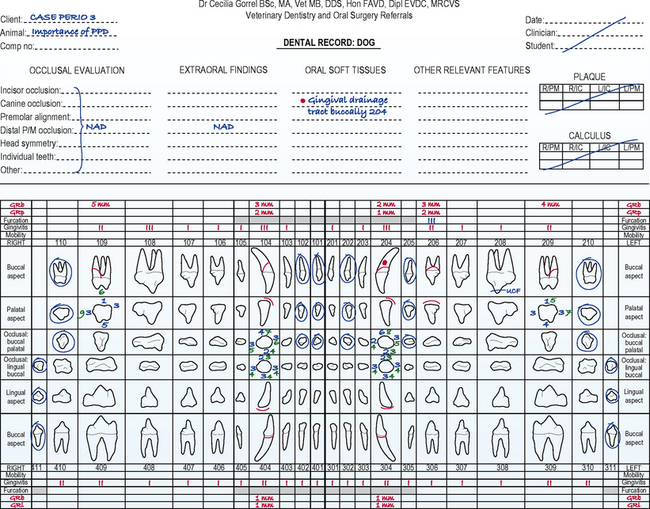
See the front page of the dental record (Fig. 8.1) for details of findings.
In summary, examination under general anaesthesia identified the following:
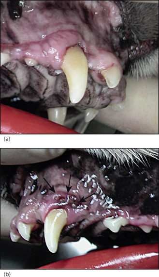
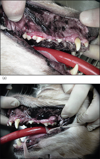
Figure 8.3 Lateral photographs centred on the right (a) and left (b) upper canine teeth.
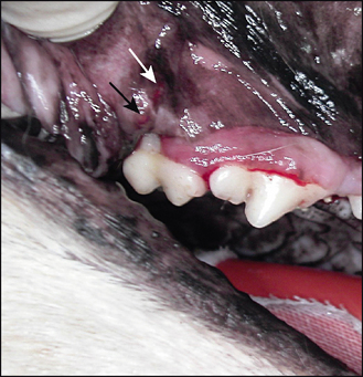
(a) The gingiva of 104 is inflamed and there is a buccal gingival recession.
RADIOGRAPHIC FINDINGS
The radiographs of 104 and 204 confirmed the clinical diagnosis of periodontitis. There was resorption of the alveolar margin and widening of the periodontal space. The radiograph of 104 showed extensive bone loss extending beyond the apex (Fig. 8.5).
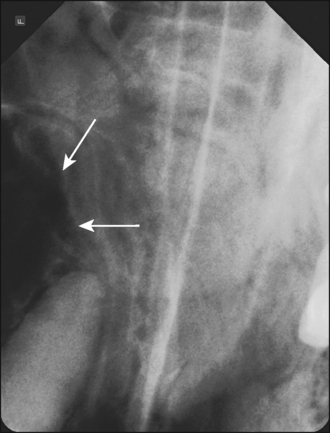
Figure 8.5 Radiograph of 104. Note the extensive bone loss on the buccal aspect that extends beyond the apex.
The radiograph of 206 showed horizontal bone loss and obvious furcation involvement (Fig. 8.6), again confirming the clinical diagnosis of advanced periodontitis.
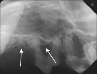
Figure 8.6 Radiograph centred on 206. Marked horizontal bone loss and furcation involvement of 206 is evident.
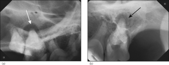
The radiographs of 109 (Fig. 8.7a) and 209 (Fig. 8.7b) demonstrated marked periapical destruction at the palatal root.
Stay updated, free articles. Join our Telegram channel

Full access? Get Clinical Tree


