Image Receptors

chapter 6 Image Receptors


GLOSSARY


Afterglow: The tendency of a luminescent compound to continue to give off light after x-radiation has stopped.


Base: A transparent flexible polyester support layer of radiographic film.


Cassette: A lightproof encasement designed to hold x-ray film and intensifying screens in close contact.


Emulsion: A layer of radiographic film made of gelatin containing suspended silver halide crystals.


Film latitude: The exposure range of a film that will produce acceptable densities.


Fluoroscopy: A special radiographic diagnostic method by which a “live view” of the internal anatomy is possible.


Intensifying screens: Sheets of luminescent phosphor crystals bound together and mounted on a cardboard or plastic base.


Latent image: An invisible image on the x-ray film after it is exposed to ionizing radiation or light before processing.


Nonscreen film: Film that is more sensitive to ionizing radiation than to fluorescent light.


Quantum mottle: An artifact of faster screens that results in density variation due to random spatial distribution of the phosphor crystals within the screen.


Reflective layer: A layer of an intensifying screen that reflects the light from the phosphor layer toward the film.


Screen film: Film with silver crystals that is more sensitive to fluorescent light emitted from intensifying screens than to ionizing radiation.


Silver halide: A compound of silver and bromine, chlorine, or iodine, all of which are in the halogen group of elements.


Supercoat: A clear protective layer on radiographic film.


INTRODUCTION


In previous chapters, discussions were limited to the production and action of x-rays. To further understand radiography, we need to discuss how a permanent record is produced using x-rays.


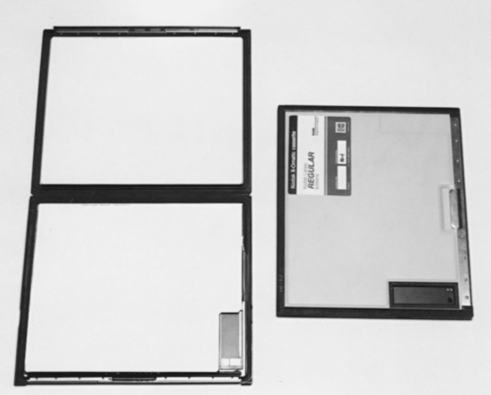
Essentially, a radiograph is formed with light-sensitive film contained in a lightproof encasement. In radiography, the lightproof encasement used most often is called a cassette (Fig. 6-1). The general-use cassette is designed to hold a piece of double-emulsion x-ray film sandwiched between two fluorescent sheets of plastic called intensifying screens. The intensifying screens are responsible for converting the x-ray radiation into visible light, which creates a latent image on the x-ray film. The film is then processed to convert the latent image into a visible image. Remarkably, more than 95% of the exposure recorded on the film is due to the light emitted from the intensifying screens. Only 5% of the exposure of the film results from the ionization of x-rays.


Many different types of image receptors and detectors convert invisible ionizing radiation into a visible image. These detectors and receptors can take many forms and, in turn, assist a number of diagnostic procedures that use radiant energy including the following:


This list is only a sample of the imaging techniques that use radiant energy. We will redirect our attention to fluorescent intensifying screens and silver halide films as image receptors.


THE CASSETTE


In radiography, the cassette is a rigid film holder designed to hold the x-ray film and intensifying screens in close contact. The cassette must be constructed with materials that are light-tight to prevent any unwanted exposure to the film yet that allow penetration of the x-rays.


The first cassettes were constructed with cardboard. This material could not be reused and thus did not pass the test of time. Over the years, cassettes made of aluminum became standard. Aluminum cassettes are still common today; however, improvements have been made to cassette fronts. As mentioned, the front of the cassette must be strong and opaque to light yet radiolucent to x-rays. Examples of available cassette fronts are those made of (1) polycarbonate (Bakelite), (2) aluminum, (3) magnesium, and (4) carbon fiber.


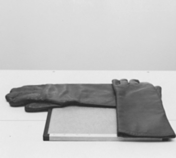
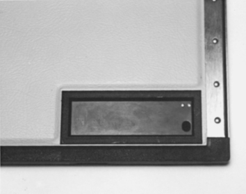
Some cassette fronts are color coded or have a colored dot on the edge to indicate the screen type inside. Color coding allows easy identification when choosing a cassette for each clinical situation. The front may also be marked into four quadrants to assist more than one exposure per film. Taking a number of exposures on one film is accomplished by exposing one quadrant while shielding the others with lead rubber strips (Fig. 6-2). An area approximately 3 × 7 cm may also be marked in the corner of the cassette front to indicate the presence of a lead blocker (Fig. 6-3). This lead blocker is present to prevent irradiation of the part of the film necessary for identification. (Film identification is discussed in Chapter 7.) Care must be taken not to superimpose any vital areas of the patient over this blocker.


The cassette front is attached to the back with hinges and catches. Several types of hinges and catches that provide a tight seal between the front and the back of the cassette are available. The closure styles range from hinges with slide catches to crossbars that pivot on a shoulder rivet in the middle of the back of the cassette. The back of the cassette is constructed with heavier material than that for the front and is normally lined with lead to absorb backscatter radiation that would cause fogging of the film.


Inside the cassette, both sides are lined with felt or foam pressure pads that ensure close contact of the film and screens. The choice of felt versus foam pads varies with each cassette manufacturer.


Cassette sizes also vary and correspond to screen and film sizes (in both metric and English). Their cost varies according to size and quality. (Note: The price quotes in most catalogs are for the cassettes only and do not include the screens.)


Cassette choice is an important aspect of veterinary radiography. The purchase of a certain cassette may help or hinder the production of quality radiographs. A cassette should have sturdy construction, maintain screen-film contact, and be user friendly in the darkroom.


Cassette Care


As with any expensive piece of equipment, the cassette should be handled with care. In veterinary medicine, cassettes tend to be exposed to some physical abuse. This is especially true in a large-animal practice. The most common causes of physical damage are (1) dropping the cassette on a hard surface and (2) leakage of fluid such as blood or urine into the cassette.


Dropping a cassette on a hard surface can result in a loss of contact between the screens and film, which results in a blurred radiographic image. (See Chapter 10 for the test procedure for screen-film contact.)


Keeping a cassette clean when working with animals is always a challenge. Precautions such as placing the cassette in a plastic bag when a “messy” situation is expected will prevent damage to the cassette’s exterior and interior. A cassette should be cleaned on a regular basis with mild soap and water. Cleaning the exterior of the cassette when the screens are cleaned (monthly) is usually adequate unless circumstances necessitate a more frequent schedule.


All cassettes should be numbered. This way, any noticeable defects on a radiograph can be traced to the “problem” cassette. Most intensifying screens within the cassette have a serial number imprinted on the screen edge. These numbers are small, however, and difficult to read. The best method of cassette identification is to number each intensifying screen near the edge or corner with a black felt-tip marker. This number will appear on each radiograph taken with that cassette. The exterior (back of the cassette) should be marked with the same number.


INTENSIFYING SCREENS


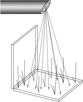
Intensifying screens are sheets of luminescent phosphor crystals bound together and mounted on a cardboard or plastic base. Two screens are normally inside the cassette to sandwich the x-ray film, which has a coating of light-sensitive emulsion on both sides (double emulsion). When the phosphor crystals in the screen are struck by x-radiation, the crystals fluoresce, and x-rays are converted into visible light (Fig. 6-4). This visible light exposes the x-ray film. As stated earlier, more than 95% of the exposure to the film is due to light emitted from the intensifying screens.


The primary purpose of the intensifying screen is to reduce the amount of radiation exposure required to produce a diagnostic radiograph. The use of screens results in lower milliamperage-seconds (mAs), thus decreasing the dose of radiation to the patient and the chance of motion on the radiograph.


Three properties determine the efficiency of the screen materials:


Screen Construction


An intensifying screen has four integral layers: (1) a base or support, (2) a reflective layer, (3) a phosphor crystal layer, and (4) a protective coat (Fig. 6-5).


The base serves as a flexible support to attach the phosphor layer to the cassette. The base must have a tough, moisture-resistant surface and not become brittle with extended use.


The reflective layer, which is attached to the base, is made of a white substance such as titanium dioxide. The purpose of the reflective layer is to reflect the light emitted by the phosphor layer back toward the x-ray film. The reflective layer increases the efficiency of the screen so that none of the light photons are lost through the base layer.


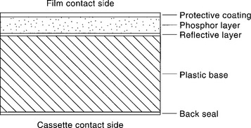
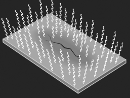
The phosphor crystal layer consists of uniformly distributed phosphor crystals held in place with a binder material. It is extremely important that this layer not change in thickness, crack, or discolor with age. Any variance in screen uniformity would alter the amount of light produced when irradiated and would alter the uniform exposure of the film (Fig. 6-6).


The protective coat is a clear coating placed on the outer surface of the screen; it provides the necessary protection to the phosphor layer. This layer must be strong enough to resist marks and abrasions and easy to clean. Veterinary radiography has many pitfalls, one of which is animal hair. Any foreign material caught in the cassette between the intensifying screen and the film will alter the exposure to the film. The debris on the screen will result in radiographic artifacts (Fig. 6-7). Because of the likelihood of artifacts and the need for subsequent screen cleaning, the protective surface must be durable and resistant to deterioration.


May 27, 2016 | Posted by in ANIMAL RADIOLOGY | Comments Off on Image Receptors

Full access? Get Clinical Tree

Get Clinical Tree app for offline access