10 William B. Thomas and Luisa De Risio The importance of the history and examination in patients with spinal pain or neurologic deficits cannot be overemphasized. At times, the diagnosis may seem straightforward, for example, when a middle-aged dachshund presents with back pain and paraparesis. But focusing prematurely on a particular diagnosis to the exclusion of other possibilities is the single most common cause of diagnostic error [1]. After all, dachshunds can suffer other causes of spinal pain or neurologic deficits. With the increasing availability of sophisticated diagnostic techniques such as magnetic resonance imaging (MRI), clinicians may wonder if the neurologic examination has become obsolete [2]. However, recommendations regarding diagnostic tests should be based on the results of the history and examination. Not every dachshund with back pain needs diagnostic imaging. Also, it is important to focus diagnostic testing on the appropriate region as determined by the neurologic examination. Imaging the thoracolumbar region in a patient with a cervical lesion would result in a missed diagnosis. Though uncommon and typically very brief in domestic animals compared to human patients, the occurrence of spinal shock could mislead the localization if not recognized. Finally, modern imaging techniques have become so sensitive that they often show incidental lesions that are not relevant to the patient’s current condition. So, accurate interpretation of diagnostic tests always requires correlation with the history and examination. Clinicians who do not specialize in neurology are often intimidated by the neurologic examination and may feel all patients with neurologic disease belong to the realm of the specialist. But most practitioners can manage many patients with spinal disease and they should know how to examine the nervous system and interpret the results. The aim of this chapter is to present a practical guide that can be applied to any patient presenting with possible spinal cord disease. This is based on an orderly process of clinical reasoning. First, determine the patient’s clinical signs by obtaining a history and performing an examination. Then interpret these signs in the context of anatomy and physiology to localize the disease to a particular region of the nervous system, the neuroanatomic localization. Next, formulate a list of possible etiologic diagnoses considering the signalment, history, and examination. Based on this list of differential diagnoses, the clinician recommends appropriate laboratory and imaging studies to confirm or exclude the diagnostic possibilities and allow appropriate treatment. The history is the most essential part of the evaluation. An accurate history allows the clinician to formulate an appropriate differential list, if not the actual diagnosis. The history also allows the veterinarian to develop empathy for the client and patient and encourages the client to develop confidence in the veterinarian. Patience, kindness, and a manner that conveys interest and compassion facilitate this process [3]. The signalment, including the patient’s age, breed, and sex, may provide clues to the diagnosis. For example, chondrodystrophic breeds are at increased risk of intervertebral disc extrusions compared to other breeds, although any breed dog and cat can suffer disc disease [4–6]. Disc herniation is uncommon in patients less than 1 year of age, and the incidence increases with age to a peak of 4–6 years for chondrodystrophic breeds and 6–8 years for other breeds of dog [4–6]. The presenting concern, also called the chief complaint, is the starting point of the diagnostic process because it allows the clinician to focus the questioning. Ask the client to describe the signs they observed that brought them to the hospital. Open-ended questions are best, such as “What seems to be the problem with Gretchen today?” You should not interrupt. Later, the clinician can augment the client’s account with specific questions. It is important to understand the difference between observations and conclusions. Consider the client’s statement, “My dog is in pain.” Pain is a purely subjective phenomenon and cannot be directly observed. Rather the client has arrived at this conclusion based on certain observations. So ask about the specific things the client has noticed that led him or her to conclude that their pet is in pain. Examples might include, “She cries when I pick her up” and “She no longer jumps on the bed.” Be aware that clients may not use words in a medically specific manner. For example, a client may say “lameness” when they are really observing paresis. Such ambiguity must be clarified early to avoid wasting time exploring an unlikely diagnosis. Once the presenting concern is clarified, explore the details. Ask when the problem started and what has changed since the onset. Anything that may have precipitated the signs such as trauma is noted. Realize that clients are prone to assume that some recent event is the cause for the pet’s current problem. Avoid the error of concluding that temporal relationships always imply a cause-and-effect relationship. Fairly trivial trauma may be coincidental or even a result of spinal cord disease rather than the cause. For example, the patient fell off the bed because of impending ataxia or paresis, and not “the patient became weak after falling off the bed,” as the client might report. In patients with substantial paraparesis or paraplegia, it is important to ask about voluntary urination. The most reliable historical evidence that the patient is able to urinate voluntarily is the observation that the pet postures and urinates when taken outside. The client may mistake finding urine in bedding or other places as evidence of voluntary urination when this is actually involuntary leakage of urine due to an overly full bladder. Review any available records from other veterinarians including results of examinations and laboratory studies. If imaging studies have been performed previously, try to obtain copies to evaluate rather than rely on written reports. After the presenting concern is characterized fully, it helps to summarize this for the client. This reassures the client that the veterinarian has heard and understood their concerns and allows the clinician to determine if there are other concerns that were not mentioned. Ask if the pet has suffered previous bouts of spinal disease, such as neck pain, back pain, or weakness. If so, find out whether the patient recovered completely so as not to confuse residual deficits with those caused by the current condition. It is important to know about any concurrent or past systemic disease. For example, a history of carcinoma suggests metastatic disease as the cause of any neurologic signs. Considering the zoonotic potential of rabies, always determine the vaccination status before handling any dog or cat with acute neurologic disease. Vaccinations against canine distemper and feline leukemia also influence the likelihood of certain neurologic diseases. Even if other medical issues are not directly related to the current problem, they might affect long-term prognosis or alter the risk of general anesthesia required for imaging studies or surgery. Review any previous or current treatments. Having the client bring in all medication bottles is helpful as they may not accurately recall drug names or dosages. Always ask specifically about over-the-counter drugs such as aspirin, acetaminophen, or ibuprofen because many clients do not consider these as “medication.” Other body systems are reviewed to identify any other abnormalities. Gastrointestinal signs are of particular importance, so ask if the client has noticed decreased appetite, vomiting, diarrhea, or blood in the feces. Patients with spinal cord disease are at increased risk of these signs due to altered autonomic function affecting the gastrointestinal tract. Additionally, these patients may have been treated with corticosteroids and nonsteroidal anti-inflammatory drugs that have gastrointestinal side effects [7–9]. The physical examination may identify a systemic disease affecting the nervous system. An example would be fever associated with meningitis or discospondylitis. An efficient approach is to incorporate the physical examination with the neurologic examination. For example, examination of the head, eyes, ears, and mouth can be performed while testing cranial nerves. Lymph nodes can be palpated at the same time the head, spine, and limbs are palpated. The skin is examined to identify any dermatitis affecting potential surgery sites when the neck and back are assessed. At the end of a thorough neurologic examination, one has to only auscult the chest, palpate the abdomen, and record temperature, pulse, and respiration to have performed a suitable physical examination. The general components of the neurologic examination are listed in Table 10.1. Every conceivable test is not necessary in every patient. Rather, the history allows the clinician to focus on the examination. A patient with spinal disease does not need an intricate evaluation of cranial nerves and mapping of sensation throughout all dermatomes. Ambulatory animals do not need to be subjected to assessment of deep pain perception. Take care when manipulating patients with potential spinal instability. For example, withhold postural reaction testing in a nonambulatory patient with recent trauma until an unstable spinal fracture or luxation has been ruled out by imaging. Flexing the neck in toy breed dogs that may have atlantoaxial instability can be disastrous. A neurologic examination form is helpful in recording the results and prompting the clinician to not miss key components. An example is provided in Figure 10.1. Table 10.1 Major components of the neurologic examination Figure 10.1 Neurologic examination form. More than any other type of examination, the neurologic examination requires patient cooperation. Sedating an unmanageable patient is a good approach for procedures such as joint palpation or chest auscultation. But sedatives and tranquilizers alter the results of much of the neurologic examination. So anything the clinician can do to foster cooperation of the patient is time well spent. Start by observing the patient while obtaining the history. Ambulatory patients are allowed to move freely about the examination room. This allows a chance to assess mental status, behavior, posture, and gait. Also many patients are calmer after they have investigated their surroundings. When it is time for the hands-on portion of the examination, gentle petting is useful and helps establish a pleasant tone. Start with procedures least likely to upset the patient and delay disagreeable tests until the end of the examination. Many patients do not like being held down for reflex testing and obviously eliciting spinal pain and testing for pain perception are uncomfortable, so save these for last. Normal consciousness implies wakefulness and awareness and is assessed by observing for response to the environment. Lethargy is characterized by inattention and decreased spontaneous activity. It can be caused by systemic illness or brain disease. With delirium, the patient is alert but overactive and responds inappropriately. A patient in a stupor is unconscious but can be aroused with stimuli, such as sound or pain. With coma, the patient is unconscious and cannot be aroused even with painful stimuli. Stupor and coma are commonly caused by a brain stem lesion or severe, diffuse disease of the forebrain. Abnormal behavior is identified by comparing the patient’s behavior to expected behavior for animals of a similar breed and age. The client is often able to bring subtle changes in behavior to the veterinarian’s attention. Behavioral abnormalities suggest a forebrain lesion. The posture should be upright with the head held straight. Animals with neck pain often carry the head lower than normal. In some cases, the thoracolumbar region is arched (kyphosis), mimicking the posture seen with back pain. Patients with thoracolumbar pain often exhibit thoracolumbar kyphosis and rigidity. With caudal lumbar or lumbosacral pain, the patient may stand with the lumbosacral region flexed and the thoracic limbs placed caudally, shifting weight cranially. A wide-based stance is common in patients with ataxia. Head tilt, where one ear is held lower than the other, usually indicates vestibular dysfunction. Head tilt must be differentiated from head turn or torticollis. A head turn is when the head is held level (one ear is not lower than the other) but the nose is turned right or left. An animal with forebrain lesions may tend to turn the head and circle in one direction. Torticollis is an abnormal curving or twisting of the neck and can occur with developmental vertebral abnormalities or spinal cord lesions that affect the gray matter or the vestibulospinal tract in the white matter [10]. Animals with severe paresis or paralysis can exhibit several different postures. Patients with mid- to caudal lumbar lesions often sit upright with their pelvic limbs extended cranially, bearing weight on their ischium. With lesions near the thoracolumbar junction, the patient sits up with the pelvic limbs placed caudally. With cranial thoracic lesions, the patient may not be able to sit upright because of paresis of the paraspinal muscles in the thoracic and lumbar region. With tetraparesis caused by a cervical lesion, the patient may be recumbent, on their side, and unable to sit up, and with severe tetraplegia there may be respiratory compromise. Acute lesions of the thoracic or lumbar segments can result in Schiff–Sherrington posture, characterized by extension of the thoracic limbs and paralysis of the pelvic limbs. There may also be extension of the head and neck (opisthotonus). Despite the increased extensor tone, the thoracic limbs are not paralyzed and exhibit normal voluntary movement. Schiff–Sherrington posture should not be confused with two other causes of increased extensor tone. Decerebrate rigidity produces extension and paresis of all limbs and opisthotonus. This posture is caused by a brain stem lesion and affected patients typically have decreased consciousness. Decerebellate rigidity occurs with acute cerebellar lesions and is characterized by opisthotonus, thoracic limb extension, and flexion or extension of the hips without paresis. In addition, animals with back pain and fear/stress associated with coming to the hospital may exhibit what appears to be extensor rigidity in the thoracic limbs, and this is often overinterpreted as Schiff–Sherrington posture. A nonslick surface, such as carpet, grass, or pavement, is used to assess gait. Observe the gait from the side, front, and rear. The patient, if able, should be walked, trotted, turned in circles, and walked up and down a short flight of stairs. Cats may be motivated to move by employing toys they can follow/chase, or by moving their carrier to different locations because cats will often want to hide back inside the carrier. Videotaping the patient and reviewing the tape in slow motion is helpful in characterizing subtle abnormalities. Each foot should come off the ground crisply with no scraping or dragging, clear the ground evenly, and land smoothly without slapping the ground. Each stride should cover approximately the same distance. Mild abnormalities are often most evident when the patient is turning. Subtle neurologic deficits are often more evident at the walk, while orthopedic disease is usually more apparent when the patient is trotting. Limb pain can cause a limp when the patient tries to bear weight briefly and gingerly on a painful limb and then quickly and forcefully plants the contralateral limb to relieve the pain. As a result, the stride of the painful limb has a shortened weight-bearing phase. When a single limb is severely painful, it is often carried. This is in contrast to a paretic limb, which dragged. Patients with bilateral limb pain, such as from hip disease or ruptured cruciate ligaments may not walk at all or have short-strided, stilted gaits, which might be confused with neurologic disease. Supporting the patient and evaluating proprioceptive positioning will often resolve this confusion. Likewise, some neurologic disorders cause lameness suggestive of orthopedic disease. For example, attenuation of a nerve root or spinal nerve by intervertebral disc extrusion often results in lameness of the limb innervated by the damaged nerve, called nerve root signature. The ability to stand and move requires intact motor and proprioceptive systems. Proprioception detects the position or movement of body parts. Receptors sensitive to movement and stretch are located in muscles, tendons, and joints. This information is conveyed by peripheral nerves to the spinal cord, which integrates local reflexes involved in posture and movement. Proprioceptive information also travels through ascending spinal tracts to the brain stem, cerebellum, and forebrain, which integrate coordinated movement. A lesion affecting the proprioceptive pathways causes ataxia. Ataxia is an inability to perform normal, coordinated motor activity that is not caused by weakness, musculoskeletal abnormality, or abnormal movements, such as tremor. The three types of ataxia are general proprioceptive, cerebellar, and vestibular. Spinal cord lesions cause general proprioceptive ataxia, characterized by a swaying, staggering gait. There is a delay in the onset of the swing phase of gait (protraction). The limb may adduct or abduct during protraction and there may be scuffing or dragging of the foot. Cerebellar ataxia is caused by cerebellar disease and is characterized by errors in the rate and range of movement, especially hypermetria. Limb movements are abrupt in onset with overflexion of the limbs on protraction. The limbs are placed inappropriately and there is swaying of the trunk and head. Vestibular ataxia is caused by a lesion in the inner ear or brain stem/cerebellum. It is characterized by leaning, falling, or rolling to one side. Other signs of vestibular disease, such as head tilt and abnormal nystagmus, may be evident. With bilateral vestibular dysfunction, the patient maintains a crouched position, is reluctant to move, and exhibits side-to-side head movements. Normal function of the motor system requires a complex interplay between the brain, descending motor tracts in the brain stem and spinal cord, motor neurons, peripheral nerves, neuromuscular junctions, and muscles. Any lesion in this system can cause paresis, a deficiency in the generation of gait or ability to support weight. Paresis is evident as a decreased rate or range of motion, increased fatigability, or inability to perform certain motor acts. Paralysis is a complete loss of voluntary motor function, while paresis indicates a partial loss of voluntary motor function. Lesions compromising the motor system affect either the upper motor neuron (UMN) or the lower motor neuron (LMN). This scheme is a physiologic one, not an anatomic one, particularly in the case of the UMN, which is functionally composed of a series of neurons extending from their origin in different regions of the brain to their termination on the LMN in the spinal cord. Disruption of the UMN pathway causes paresis or paralysis, normal or exaggerated spinal cord reflexes, and normal or increased muscle tone (spasticity). With spinal cord disease, UMN paresis is compounded by ataxia. When walking, the combination of a delay in the onset of protraction, overextension of the limb in protraction and spasticity result in an increased stride length and a “floating” gait. This is distinct from the overflexion of the limb seen with cerebellar lesions.
History, Neurologic Examination, and Neuroanatomic Localization for Spinal Cord and Nerve Root Disease
History
Signalment
Presenting concern
Past medical history
Physical examination
Neurologic examination
Mental status and behavior
Posture
Gait
Postural reactions
Spinal reflexes and muscle tone
Cranial nerves
Palpation
Sensory testing 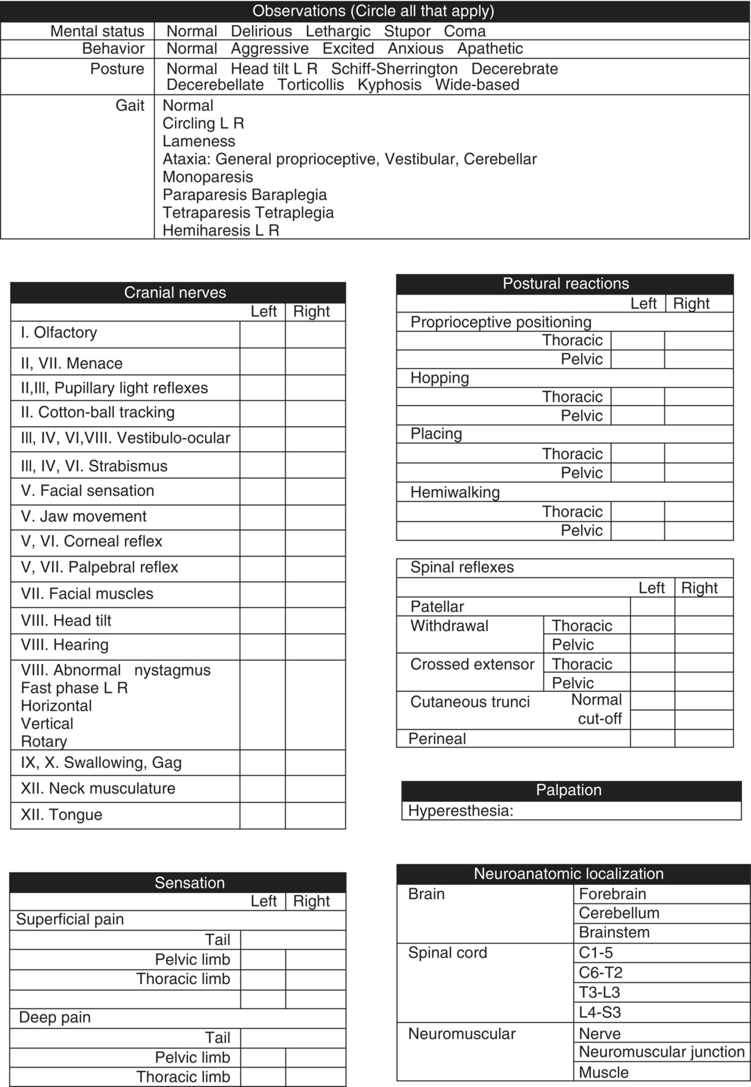
Mental status and behavior
Posture
Gait
Stay updated, free articles. Join our Telegram channel

Full access? Get Clinical Tree


