49 Histiocytoma
INTRODUCTION
Canine cutaneous histiocytoma is a common benign solitary tumour that usually undergoes spontaneous regression. They tend to occur in younger dogs, on the pinnae, face or distal extremities. Histiocytoma can be difficult to differentiate from other tumours, such as plasmacytoma or mast cell tumour, and from localized granulomatous infections, just on clinical presentation alone. A systematic approach is required to make a diagnosis and then to taking the appropriate action, which will depend on the individual assessment.
CASE HISTORY
The tumour has usually been noticed by the owner prior to presentation, although it may be detected on veterinary examination of the dog for other reasons. Dogs may draw their owner’s attention to the tumour by obsessive licking. There are no systemic signs associated with histiocytoma.
In this case a small mass on the dog’s right forelimb was noticed by the owner about 10 days prior to presentation. The dog had started to lick it and its size had increased. The dog’s general health was unaffected.
CLINICAL EXAMINATION
Histiocytoma appears as a small (1–3 cm), solitary circumscribed dome or button and may, by the time of presentation, have an ulcerated surface with haemorrhagic crusts. The tumour is most commonly located on the face, pinnae or limbs and some dogs may have lymphadenopathy of the draining lymph node.
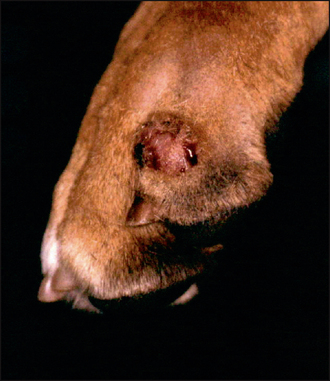
In this case a small, dome-shaped tumour with haemorrhagic surface crust was present on the lateral aspect of digit 5 of the left forefoot (Fig. 49.1). The draining lymph node was not palpable.
CASE WORK-UP
The history (age, breed and duration of onset) and clinical examination were suggestive of a cutaneous histiocytoma; however, the other differentials could not be ruled out on clinical examination alone. Boxers in particular are predisposed to mast cell tumour, so confirmation of the diagnosis of histiocytoma was essential, before deciding on the best course of treatment.
A fine-needle aspirate sample was obtained aseptically with a 23-gauge needle. The author’s technique is to insert the needle into the lump and redirect it in and out in several directions and then remove it. A 10-ml syringe containing air is attached to the needle and the contents expressed onto a clean microscope slide by pushing in the plunger. Usually, this method produces a good harvest of cells. The slide is stained with modified Wright’s stain or sent to a cytopathologist. A correct interpretation of the sample is essential to avoid a misdiagnosis, which could lead to major surgery or to other life-threatening treatments.
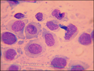
In this case, the fine-needle aspirate revealed a population of mononuclear cells with oval to round nuclei (some with indented nuclear membranes) and pale blue cytoplasm (Fig. 49.2). The cells lacked the granules that one would expect to see with mast cells, thus ruling out a mast cell tumour. Plasmacytomas have a smooth surface that is rarely ulcerated. Cells from a plasmacytoma generally have a more basophilic staining cytoplasm, and the cells show increased anisocytosis (variation in cell size) and anisokaryosis (variation in the size of the nucleus). Sebaceous gland adenomas and hyperplasia yield epithelial cells containing lipid vacuoles, which were not seen in this case, and infectious granulomas would yield large numbers of inflammatory cells such as neutrophils and macrophages that also were not present in this case.

Figure 49.2 Round cells with round or indented nucleus and pale blue staining cytoplasm (note lack of granules).

Stay updated, free articles. Join our Telegram channel

Full access? Get Clinical Tree

