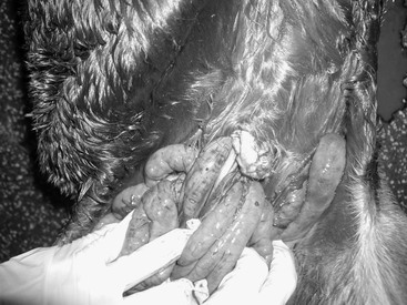
Laura A. Werner Umbilical hernia has a reported incidence of 0.5% to 2%, and has even been reported as high as 29.5%, making it the most common type of hernia observed in foals. Anatomically, this type of hernia consists of intact peritoneum covered with fascia and skin, and a defect at the linea alba where an umbilical scar has failed to form. Umbilical hernias are congenital and are often associated with trauma to or pressure on the umbilicus during birth or with umbilical infection. In severe cases of umbilical trauma, evisceration can occur through the umbilicus immediately after parturition (Figure 179-1). This is an immediate surgical emergency necessitating prompt referral and repair; a successful outcome can be expected as long as the intestine and mesentery have not been unduly compromised. Most umbilical hernias less than 2 cm in diameter will resolve spontaneously if they are repeatedly manually reduced. Repair of some smaller hernias, or hernias larger than 4 cm, is performed for cosmetic reasons or to prevent possible incarceration of the intestine. Banding or clamping of smaller hernias is commonly performed. Clinical signs such as sudden enlargement of the hernia, ventral edema, signs of colic, and inability to easily reduce the hernia all can be indications of a surgical emergency. A large plaque of ventral edema is commonly a sign that omentum has been incarcerated in the hernia. Umbilical hernia may or may not be associated with signs of colic. Evagination of the urinary bladder through an umbilical hernia also has been reported. Parietal or Richter’s hernias can also occur, with the antimesenteric surface of the bowel, most commonly the ileum, becoming entrapped in the umbilical hernia. Partial obstruction or complete obstruction, as well as necrosis of the bowel, can occur. Large and small colon can also be involved with parietal hernias. Enterocutaneous fistulas can develop in chronic parietal hernias. Ultrasound examination of the hernia is helpful in determining its contents and viability of incarcerated bowel. Correction of umbilical hernia can be achieved in uncomplicated cases by placement of clamps, elastrator bands, or hernia belts. The clamps or bands are generally placed with the foal in dorsal recumbency and under short-term general anesthesia. Complications of the use of clamps or bands can include colic, abscess formation, dislodging of the bands or clamps, and parietal hernia. A reported complication rate of 19% was reported in one study in which 40 cases were evaluated. An equine hernia belt applied for several weeks can also be used in foals to resolve a small hernia. Hernias less than 6 cm (three or four finger widths) in length in foals younger than 6 months respond best to the latter treatment. A hernia belt is often chosen because the treatment cost may be less than that associated with surgical repair. Surgical repair of umbilical hernias can be performed by open or closed herniorrhaphy techniques. Surgery is recommended if the foal is older than 6 months of age, for hernias longer then 6 to 8 cm, or whenever intestine or omentum is incarcerated in the hernia. The foal is anesthetized, and the area around the hernia is surgically clipped and aseptically prepared. An elliptical incision is made in the skin around the hernia sac and is continued down to the level of the hernia sac. The hernia sac can be inverted or removed. Preplacement of interrupted sutures in a tension-relieving pattern, such as vertical mattress, near-far-far-near, or vest-over-pants pattern, with an absorbable suture material is recommended. The subcutaneous tissue and skin are then closed in a routine manner. Perioperative broad-spectrum antimicrobials are recommended. Stall rest for 30 days is recommended to prevent failure of the repair. Colic, edema at the incision site, dehiscence, and pneumonia have all been reported as complications of herniorrhaphy. Complication rates have been reported to be similar to those associated with clamping techniques overall, although the rates are higher for complicated umbilical hernias. Diaphragmatic hernias in the foal can be congenital or may arise secondary to trauma during parturition. They can also often develop secondary to rib fractures in the foal, especially when fractures involve ribs 3 through 8 at the costochondral junctions. In recent reports involving 31 cases of diaphragmatic hernias, 6 cases were foals younger than 1 year. Colic was the presenting complaint in all cases. Diagnosis is often made on the basis of several imaging modalities, and the smaller the size of the foal, the easier the diagnosis can be made with ultrasound and radiography. Hemothorax, hemoabdomen, or both may be seen in neonates with diaphragmatic hernia. Respiratory distress is another common presenting sign in foals. The small intestine is most frequently herniated through the defect, but the large intestine, stomach, liver, and spleen can also be involved. Surgical repair can be aided by placing the foal in reverse Trendelenburg position to help the viscera fall away from the defect and facilitate repair. Positive-pressure ventilation is necessary during anesthesia. From an abdominal approach, the viscera can be retrieved from the thorax, and the defect can be closed by suturing alone or by both suturing and stapling a mesh over the defect. Hernias involving the ventral portion of the diaphragm are more amenable to repair than hernias involving the dorsal aspect, near the ribs. Thoracoscopic approaches or rib resection techniques have been reported for repairing defects in the dorsal part of the diaphragm. Evacuation of the air in the pleural cavity is necessary after repair, and an indwelling chest tube may be placed for additional evacuation of pleural cavity air during recovery or after surgery. Rib fractures can be repaired concurrently with a combination of surgical plate and wire techniques to prevent further trauma to the diaphragm or thoracic cavity contents. The prognosis for survival is guarded (46% survival rate). Foals can die or undergo euthanasia because of the severity and extent of intestinal compromise. Foals that survive through the surgery may die during recovery.
Hernias in Foals
Umbilical Hernia

Treatment
Diaphragmatic Hernia
Treatment
![]()
Stay updated, free articles. Join our Telegram channel

Full access? Get Clinical Tree


