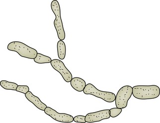
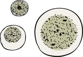
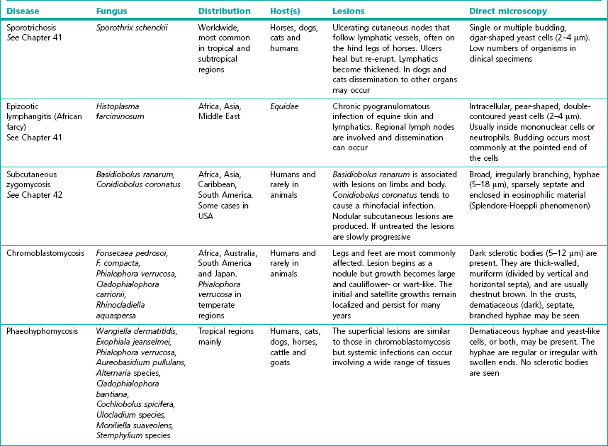
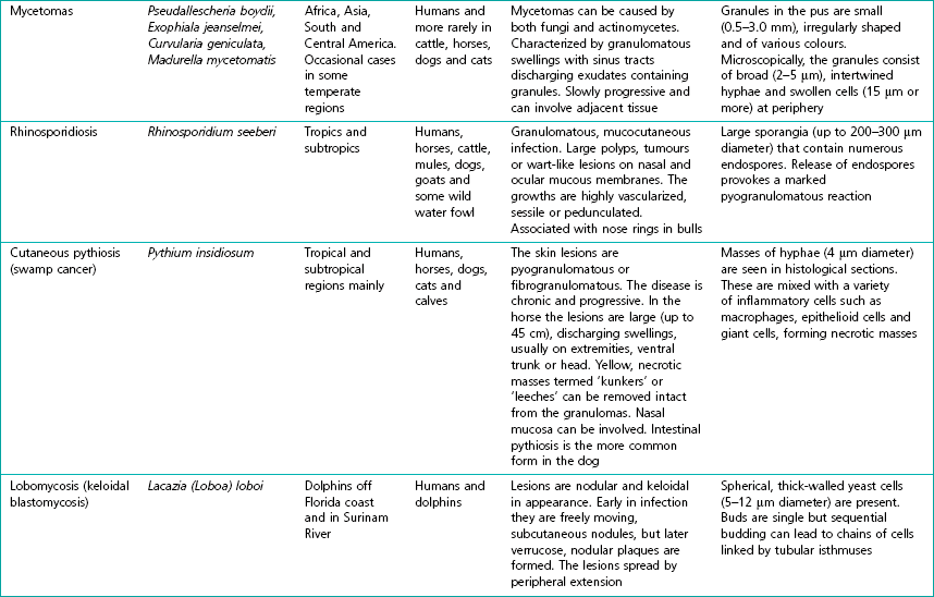
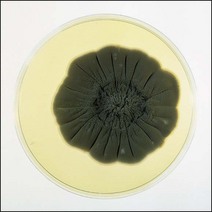
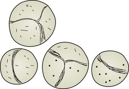
Chapter 43 The subcutaneous mycoses are all transmitted in a similar manner. Fungi that are normally saprophytes in soil, vegetable debris, water or on plants become implanted in the skin due to trauma, with the subsequent development of a subcutaneous infection that is usually chronic. Occasionally there is spread of the infection with involvement of other organs. With the exception of sporotrichosis, which occurs worldwide, the subcutaneous mycoses are most common in tropical and subtropical regions. Table 43.1 summarizes the subcutaneous mycoses and gives the causative fungi, distribution, main hosts, type of lesions produced and the appearance of the fungi on direct microscopy of the specimen. The phaeoid fungi involved in chromoblastomycosis and phaeohyphomycosis are dematiaceous or darkly pigmented due to the presence of melanin in their hyphal walls (Fig. 43.1). The actual fungal species isolated from a lesion often reflects its relative abundance in the particular geographical area. For example, Phialohora verrucosa is the most common cause of chromoblastomycosis in humans when the condition occurs in temperate regions. Dark sclerotic bodies (5–12 µm in diameter) are characteristic of chromoblastomycosis. These brown, thick-walled, multiseptate forms known as muriform bodies (sclerotic cells, Medlar bodies) are thought to represent an intermediate vegetative form, phenotypically arrested between a yeast and a mould (Fig. 43.2). The lesions of phaeohyphomycosis contain dematiaceous hyphae and yeast-like cells but no sclerotic bodies (Fig. 43.3). Chromoblastomycosis occurs uncommonly in humans, toads and frogs and is extremely rare in other animals. Phaeohyphomycosis occurs sporadically in cats, dogs, horses, cattle and goats, sometimes as a disseminated infection. Figure 43.3 Dematiaceous hyphae (5–10 µm in diameter) as seen in an aspirate from a case of phaeohyphomycosis. • Rhinosporidium seeberi, causing rhinosporidiosis, has been cultured in vitro using a human rectal tumour cell line. The normal habitat of this novel aquatic protistan parasite is thought to be stagnant water. It is associated with chronic, polypous rhinitis characterized by the presence of large sporangia (spherules) containing numerous endospores in affected tissues (Fig. 43.4). Figure 43.4 Sporangia of various sizes (6–300 µm in diameter) that occur in nasal polyps in rhinosporidiosis.
Fungi causing subcutaneous mycoses
![]()
Stay updated, free articles. Join our Telegram channel

Full access? Get Clinical Tree


