Functional Anatomy of the Palmar Aspect of the Foot

Chapter 29 Functional Anatomy of the Palmar Aspect of the Foot


The palmar portion of the foot consists of several important structures functioning to support the foot and the limb of the horse, as well as being an integral part of the energy dissipation mechanisms present within each limb. These palmar foot structures include the cartilages of the foot (also called the lateral collateral or ungual cartilages), the digital cushion, the frog, and an extensive vascular network. Although each of these structures is present in every foot, morphological features and tissue composition vary widely among horses, which may be responsible for differing efficiencies in ability to dissipate energy. Furthermore, such differences may in part account for differences between the feet of a sound horse and the feet of a horse with chronic lameness associated with the foot. Awareness of how these tissues interact and relate to one another during foot impact is important for understanding how the foot dissipates energy and how potential problems may arise to produce lameness when energy dissipation is not efficient and the concussive forces are transmitted to the bones and other connective tissues. The domestic horse spends considerable time standing, so the structure of the palmar aspect of the foot is important for support to the weight of the horse shifted dorsally toward the connective tissues at the toe of the foot.


The medial and lateral cartilages of the foot extend from the palmar surface of the distal phalanx to the bulbs of the heel as large vertical sheets, whereas the digital cushion lies between the medial and lateral cartilages of the foot and extends dorsally toward the solar surface of the distal phalanx distal to the deep digital flexor tendon (DDFT). Associated with each cartilage of the foot is a venous network that connects with the venous vessels under the distal phalanx and the vessels associated with the dermis of the hoof wall. The venous microvasculature forms a hydraulic system that is hypothesized to provide the mechanism for how the ground impact energies are dissipated, before these forces are transmitted to and damage the bone and other connective tissues within the foot. Horses with good or excellent hydraulic systems should be more efficient in dissipating the impact energy compared with horses with feet with less-well-developed hydraulic networks.


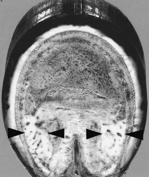
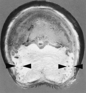
The cartilages of the foot lie beneath the skin and dermis and the coronary venous plexus and have previously been described as rhomboid-shaped, with a convex surface abaxially and a concave surface axially. Several ligaments secure the cartilages of the foot to the digital bones.1-4 However, these descriptions are overly simplistic and appear to have been obtained from examination of horses with underdeveloped structures of the palmar aspect of the foot. The morphological features of the cartilages of the foot vary greatly, with a range of shapes and thickness, the presence of an axial projection from its distal edge, and the extent of vascularity.5 The structure of the cartilages of the foot is best determined by viewing the foot in transverse sections (Figures 29-1 and 29-2). In frontal (dorsal) sections cut perpendicular to the ground, beginning at the bulbs of the heel, the cartilages of the foot have a C– to L-shaped configuration. Both the upright and the base parts of the L-shaped cartilage vary in thickness among horses. The mean thickness of the upright part at the level of the navicular bone ranges from 0.5 to 2.0 cm in an adult horse (450 to 550 kg body weight). The base part or axial projection of the L-shaped cartilage varies in its thickness and the distance that it extends toward the midline of the foot overlying the bars and the frog. The cartilages of the foot are thinnest in the heel region (0.45 to 1.3 cm) but become thicker closer to the distal phalanx (0.6 to 1.5 cm) and thin slightly as they attach onto the distal phalanx (0.5 to 1.0 cm). The cartilages of the foot are thicker in forelimbs than hindlimbs.


Overlying the abaxial surface of the cartilages of the foot are a plexus, the laminar dermis, and the hoof. Loose connective tissue extends from each cartilage to the DDFT. On the axial surface the axial projection of the cartilage extends toward the midline of the foot. In most feet, this axial projection extends from the dorsal half of the cartilage; the palmar half of the cartilage has virtually no, or very small, extension into the substance of the digital cushion. However, in some feet it extends the entire dorsal-palmar extent of the cartilages of the foot. This axial projection extends to overlie the epidermal ridges of the bars, with many fingerlike projections extending into the substance of the digital cushion, and may extend across the midline of the palmar aspect of the foot under the digital cushion to fuse with that of the opposite side of the foot. These white bundles of fibrous and fibrocartilaginous tissues are easily discerned from the surrounding yellow elastic, adipose, and collagen fibers of the digital cushion. The relative thickness of the axial projection in a distal-proximal orientation varies. In horses younger than 4 to 5 years of age the axial projection usually is not fully developed along the entire length of the cartilage, and in young foals there is only a thin sheet of fibrous tissue.


The cartilages of the foot contain primarily hyaline cartilage, but in many horses from 4 to 5 years of age the medial border of the cartilage develops fibrocartilage. A fibrocartilaginous ligament of variable thickness develops between the cartilage and the DDFT; this is consistently larger in forelimbs than hindlimbs. In some horses the cartilages of the foot can ossify. This may be genetically controlled, but any stress on the foot, that accentuates vibration and results in higher energy forces being transmitted through the foot, may promote ossification.


Several ligamentous attachments connect the cartilages of the foot to the distal and middle phalanges, as well as to the navicular bone.3,4 The chondroungular ligaments attach the cartilage to the distal phalanx along the palmar process, whereas the medial and lateral chondrocoronal ligaments attach the cartilage to the proximal half of the middle phalanx. The medial and lateral ligaments of the cartilages of the foot (collateral chondroungular ligaments) attach the cartilage to the angle of the distal phalanx. The paired chondrosesamoidean ligaments attach the axial surface of the cartilage to the navicular bone. A pair of elastic ligaments extends between the proximal phalanx and the proximal surface of the cartilages of the foot; these ligaments are most prominent in larger horses, such as draft breeds. Cruciate ligaments of the cartilages of the foot (cruciate chondroungular ligaments) connect the axial surface of the cartilage to the palmar process on the opposite side of the foot. Within the digital cushion are fiber tracts radiating from the connective tissue ventral to the attachment of the DDFT (digital torus), through the digital cushion, to the axial surface of the cartilages of the foot.

Stay updated, free articles. Join our Telegram channel

Jun 4, 2016 | Posted by in EQUINE MEDICINE | Comments Off on Functional Anatomy of the Palmar Aspect of the Foot

Full access? Get Clinical Tree

Get Clinical Tree app for offline access