Chapter 59Flexural Limb Deformities in Foals
Congenital Flexural Deformities
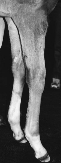
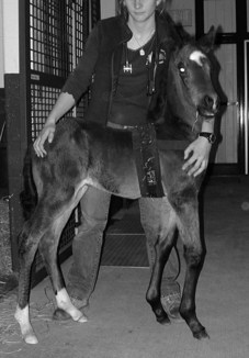
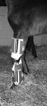
Mild flexural deformities of the carpal, metacarpophalangeal or metatarsophalangeal, or DIP joints resolve spontaneously if the foal has the ability to stand, nurse, and ambulate on its own. Most foals with mild and moderate deformities of these sites respond favorably to physiotherapy, by manually extending the limbs every 4 to 6 hours for 15-minute sessions, or forcing the foal to ambulate (Figure 59-1). Heavy bandaging, splinting or casting, and the administration of oxytetracycline can be helpful.2,8 Foals with deformities advanced enough to require assistance to stand respond more rapidly after application of a cast, which should be changed every 2 to 3 days (Figure 59-2). If a carpal deformity severe enough to prevent standing is recognized at parturition, the foal should be heavily sedated, within 30 to 45 minutes, and full-leg casts should be applied with the limbs placed in extension. These should be changed within 24 hours and reset. Generally a rapid response is seen after the first or second application of casts. If the foal develops any complications with the casts or becomes distressed, casts should be removed. Commercially available articulating braces (Almanza Corrective Boot, www.redboot.com.ar/home.html; Equine Bracing Solutions, Trumansburg, New York, United States; Dynasplint Systems, Serverna Park, Maryland, United States) are available and allow adjusting the degree of extension in a specific region of the limb (Figure 59-3). As with all splints, caution must be exercised to prevent pressure sores. It is important to assist the foal in nursing and to provide supportive care in an intensive neonatal facility, because these horses are at high risk for developing complications with other body systems.

Fig. 59-1 Mild bilateral flexural deformity of the carpus. This degree of deformity is self-limiting.
Stay updated, free articles. Join our Telegram channel

Full access? Get Clinical Tree


