CHAPTER 53Fetal Sex Determination
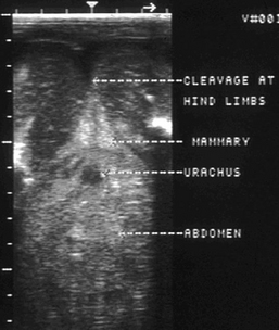
Equine fetal sexing is the determination of sex of an equine fetus after 55 days of gestation. Different techniques for determination are required for the different stages of gestation. Technique 1, performed between 55 and 90 days of gestation, involves finding the genital tubercle (precursor to the penis in the male and precursor to the clitoris in the female) and determining its location relative to other fetal structures. The genital tubercle appears as a hyperechoic bilobulated “equals” sign 2 to 3 mm in length and develops between the hind legs on the ventral midline of both sexes at 50 to 55 days. Migrations of the tubercle that indicate the sex of the fetus begin about day 54 to 55. A determination cannot be made before 55 days because the migration toward the umbilical cord in the male or toward the anus in the female has not yet occurred. Using technique number one, the veterinarian should be able to make a diagnosis 95% of the time on the first examination with 99% accuracy. The examination should take from a few seconds to 5 minutes to perform.
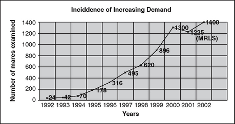
If a practitioner is the only one in his or her area able to accurately perform a sexual determination, the procedure can develop into a new worthwhile profit center for the practice. Figure 53-1 is a graph of increasing demand when fetal sex determination was introduced into the area.
MATERIALS NEEDED FOR FETAL SEX DETERMINATION
FETAL DEVELOPMENT
See Tables 53-1 and 53-2 and Figure 53-2.
Table 53-1 Fetal Anatomical Development Chart
| Days | Visualization |
| 55-60 | Fetus is very small; genital tubercle is difficult to see and may or may not be fully migrated. |
| 60-70 | Fetus is easily accessible; tubercle is distinct and fully migrated. (Easiest and most ideal time for fetal sex determination) |
| 70-80 | Fetal tubercle is distinct but slightly more difficult to reach. |
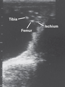
| 80-90* | Difficult to access fetus; tubercle is less distinct; genitalia development is just beginning. (Most difficult time) (Figure 10-2) |
| 90-100 | Fetus is generally accessible; genitalia are not very well developed. (Difficult to differentiate genitalia at times) |
| 100-110 | Genitalia becoming more evident. |
| 110-120 | Fetus is very accessible, and genitalia is well developed. (Ideal time in technique 2 for determination) |
| 120-140 | Genitalia are well developed, but posterior of fetus may be difficult to access at times. |
| 140-150† | At times fetus has anterior presentation with posterior out of reach. |
| 150+ | Usually the fetus has anterior presentation, and posterior of fetus is out of reach. Transrectally this stage is difficult, with a low percentage diagnosis of 5-25%. |
| 150 to Term | Transabdominal—low percentage diagnosis, time consuming, need to clip abdomen, more powerful ultrasound machine is helpful. |
* At 80 to 90 days the fetus is often difficult to reach due to the position of the uterus in the posterior abdomen. At approximately 80 days the allantoic fluid of the pregnancy pulls the uterus over the rim of the pelvis. The fetus is small, falls to th e most ventral portion of the uterus, and is very difficult to reach. As the uterine contents (fetus and fluids) increase in size, the uterus actually elevates more in the abdominal cavity and becomes easier to reach and view the fetus.
† Mares that are 130 to 150 days that are out of reach should be viewed again for possible position change.
Table 53-2 Ultrasonic Cross Sectional Anatomy Found in Planes I, II, and III
Stay updated, free articles. Join our Telegram channel

Full access? Get Clinical Tree


