36 Feline constipation and megacolon
Initial presentation
Tenesmus, haematochezia and abdominal distension
Signalment: 8-year-old, female neutered Devon Rex cat, body weight 2.5 kg
Case history
The cat was presented for tenesmus, haematochezia and abdominal distension, accompanied by anorexia and lethargy. Since being acquired 4 years earlier she had had intermittent episodes of constipation, manifested as straining to pass hard faeces which were occasionally coated with fresh blood or mucus. The episodes had initially occurred two or three times per year, but for the past 6 months had been occurring monthly. During these episodes the cat would defecate outside the litter tray and she appeared to have abdominal bloating. Symptomatic treatment with laxatives generally improved the condition within 1 to 2 days.
Occasional episodes of small volume diarrhoea were reported (once to twice a year). At the time of presentation, she had passed no faeces for 4 days and had had a poor appetite for 2 days.
The cat was acquired from a rescue centre 4 years previously and lived indoors with one other unrelated Devon Rex cat. She was vaccinated against feline herpesvirus-1, calicivirus and feline parvovirus and de-wormed every 6 months with praziquantel and pyrantel. The cat was fed a proprietary canned cat food.
She was thought to have previously been used for breeding and was reported to have had surgery for an intussusception at 2 years of age.
Physical examination
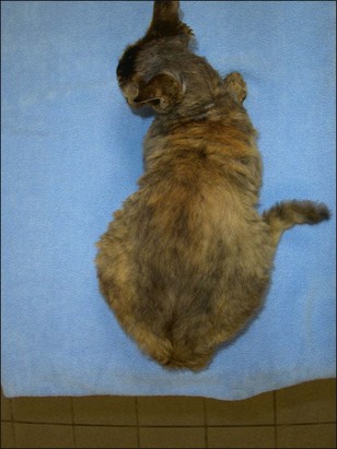
The cat appeared slightly thin (body condition score 3/9) and had a distended abdomen (Fig 36.1). Her hydration appeared adequate and mucous membrane colour was pink with a capillary refill time of 2 seconds. Thoracic auscultation revealed a Grade I to II/VI systolic murmur, with point of maximal intensity at the left sternal border. No pulse deficits or arrhythmias were detected. Respiratory rate was mildly increased at 30 breaths per minute, attributed to stress, as no abnormal lung sounds were detected. Abdominal palpation revealed a large colon, filled with hard faeces and a moderately distended bladder.
Problem list and discussion of problems
The cat’s primary problem was thought to be constipation, based on the history and physical examination findings.
Differential diagnosis
Differential diagnoses for constipation include:
With the chronic history and the lack of clinical signs consistent with most of the other possible differential diagnoses for constipation, the most likely cause in this cat was thought to be idiopathic megacolon, although other neurological causes and neoplasia could not be ruled out
Case work-up
A neurological examination was performed with no abnormalities detected. A rectal examination showed no evidence of herniation, anal sac disease, strictures or foreign bodies. A few small pieces of hard dry faeces were present in the rectum.
Minimum database
Routine haematology showed a moderate neutrophilia (26.2 × 10⁹/l; reference range 2.5–12.8 × 10⁹/l). This most likely represented a non-specific inflammatory response. Serum chemistry parameters were within the reference ranges.
Imaging
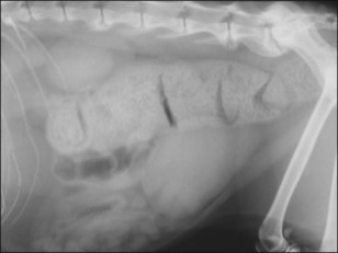
Abdominal radiography showed impaction of the transverse and descending colon with faeces (Fig 36.2). In addition, ventral spondylopathy was present at the lumbosacral junction. Two views were taken to fully evaluate narrowing of the pelvic canal.

Fig 36.2 Lateral abdominal radiograph showing impaction of the transverse and descending colon with faeces at initial presentation.
Further investigations that could have been beneficial include urinalysis for assessment of hydration, ultrasonography if intraluminal or extraluminal masses were suspected, colonoscopy if intraluminal disease is suspected and barium enema if a stenotic lesion was suspected. If the neurological examination had been abnormal, further evaluation with MRI may have been useful.
Diagnosis
A diagnosis of colonic impaction was made. A diagnosis of obstipation could not initially be made, as it was not known whether the condition was refractory to therapy. Initial management was commenced with manual evacuation of faeces with the cat anaesthetized and a warm water enema was given.
Oral therapy with lactulose (initial dose 0.5 ml po q 8 hours) and cisapride (1.5 mg/kg po q 12 hours) was then started and the owner advised to feed a low residue diet. The clinical signs improved initially, although the frequency of defecation remained decreased, occurring only every second day.
Follow-up
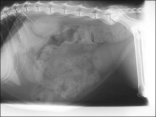
Over the following 18 months the cat required increasing doses of lactulose and cisapride and micro-enemas with sodium alkylsulphoacetate on three occasions. The cat subsequently presented after having not defecated for 4 days. She also again had a poor appetite and was lethargic. Further radiographs were obtained which showed a grossly dilated impacted colon (Fig 36.3) and as the condition was no longer responding to management a diagnosis of idiopathic megacolon was made.

Full access? Get Clinical Tree

