Chapter 6 Familial Renal Diseases of Dogs and Cats
Introduction and Pathophysiology
A. Most familial renal diseases result in chronic renal failure (CRF) at a young age (<5 years) but some are characterized by renal tubular defects (e.g., Fanconi syndrome in the Basenji) or morphologic abnormalities that result in hematuria (e.g., renal telangiectasia in Pembroke Welsh corgi dogs).
B. A familial disease is one that occurs in related animals with a higher frequency than would be expected by chance.
C. Congenital diseases are present at birth and may be genetically determined or result from exposure to adverse environmental factors during development.
D. In many familial renal diseases of dogs, the kidneys are thought to be normal at birth but undergo structural and functional deterioration early in life.
E. Some familial renal diseases of dogs probably are examples of renal dysplasia.
1. The term renal dysplasia refers to disorganized development of renal parenchyma due to abnormal differentiation and is characterized by the presence of structures in the kidney inappropriate for the stage of development of the animal.
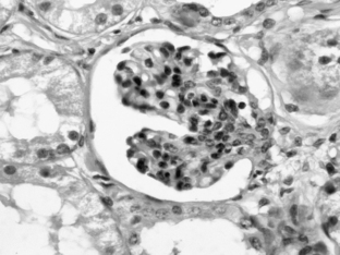
2. Lesions suggestive of renal dysplasia include asynchronous differentiation of nephrons (indicated by persistence of immature or “fetal” glomeruli; Figure 6-1) and persistent mesenchyme (usually in the medullary interstitium).
F. Many familial renal diseases are very variable in severity and rate of progression among individual animals.
G. Most of these diseases are progressive and ultimately fatal, and therapy usually is limited to conservative medical management of CRF.
Signalment
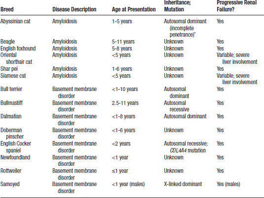
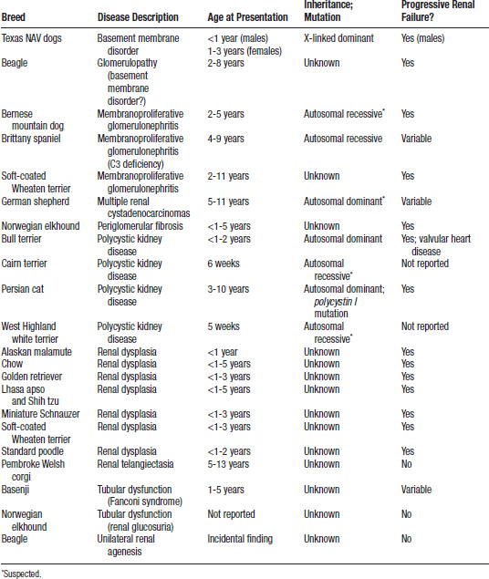
A Familial renal disease has been reported in many breeds of dog (Table 6-1) and may occur sporadically in mixed breed animals.
B. The clinician should consider the possibility of familial renal disease whenever CRF occurs in immature or young adult animals.
D. The age of onset of familial renal disease usually is 6 months to 5 years, with many animals presented before 2 years of age.
1. Renal amyloidosis in beagles and English foxhounds, suspected glomerular basement membrane disease in beagles, and telangiectasia of the Welsh corgi, however, occur in older dogs (>5 years).
History
B. Clinical findings in Basenji dogs with Fanconi syndrome may include polyuria, polydipsia, weight loss, dehydration, and weakness.
C. Hematuria, dysuria, and apparent abdominal pain may be reported by owners of Pembroke Welsh corgi dogs with renal telangiectasia.
Physical Findings
F. Small, irregular kidneys (Cairn and West Highland white terriers and Persian cats with PKD are exceptions and have kidneys that often are markedly enlarged).
G. Signs of fibrous osteodystrophy (e.g., rubber jaw) can sometimes be detected on physical examination of young growing dogs with CRF. Signs of renal osteodystrophy rarely are apparent in older dogs with CRF.
Laboratory Findings
Urinalysis
2. Proteinuria in animals with glomerular disease.
b. Abyssinian cats and Shar pei dogs with familial amyloidosis if there is sufficient glomerular involvement.
4. Hematuria in Welsh corgi dogs with renal telangiectasia or German shepherd dogs with multifocal renal cystadenocarcinomas.
Imaging
A. In many dogs and cats with familial renal disease the kidneys are small on radiographs and show irregular contour, increased echogenicity, and decreased corticomedullary distinction on ultrasound examination.
B. Renal cysts in animals with PKD have a characteristic ultrasonographic appearance (i.e., multiple round anechoic lesions with distal acoustic enhancement in both kidneys).
Pathologic Findings
A. Familial renal disease is characterized by the presence of primary dysplastic lesions, compensatory lesions, and degenerative lesions.
B. In many cases, the secondary degenerative lesions overshadow the underlying primary dysplastic lesions, making the correct diagnosis difficult.
C. Primary dysplastic lesions that have been observed in some familial renal disease of dogs include:
D. Primary dysplastic changes are most prominent in the Lhasa apso, Shih tzu, soft-coated Wheaten terriers with renal dysplasia, standard poodle, chow, and miniature Schnauzer.
E. Juvenile renal diseases in the Samoyed, English Cocker spaniel, and bull terrier result from abnormalities of type IV collagen in glomerular basement membranes and represent animal models of X-linked dominant, autosomal recessive, and autosomal dominant hereditary nephritis in humans, respectively.
Abyssinian Cat (Amyloidosis)
Clinical Course
A. Amyloid deposits first appear in the kidneys between 9 and 24 months of age and, in many cats, amyloid deposition leads to CRF within the first 3 years of life.
B. Amyloid deposition in the kidney may be mild, and some affected cats may live to an advanced age without detection of their amyloid deposits.
Pathologic Findings
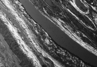
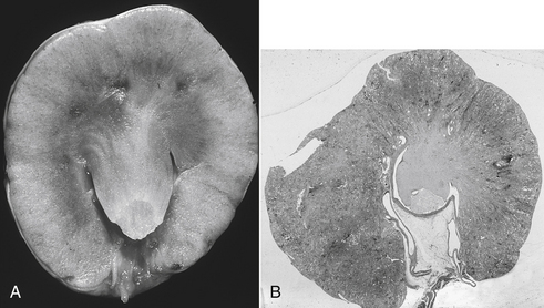
A. The principal pathologic lesions in the kidneys of Abyssinian cats with familial amyloidosis are medullary amyloid deposits (Figure 6-2), papillary necrosis (Figure 6-3), chronic tubulointerstitial nephritis characterized by lymphoplasmacytic infiltration and fibrosis, and variable glomerular amyloid deposits.
B. Glomerular amyloidosis is mild and often difficult to detect in many affected cats, but occasionally it can be severe.
C. Medullary amyloid deposition was found in all affected Abyssinians whereas glomerular deposits were found in 75%.
D. Medullary interstitial amyloid deposits interfere with blood flow to the renal papilla, resulting in papillary necrosis and secondary interstitial medullary fibrosis and mononuclear inflammation.
E. Amyloid deposition is not restricted to the kidneys in Abyssinian cats with amyloidosis, and deposits frequently are found in other organs (e.g., adrenal glands, thyroid glands, spleen, stomach, small intestine, heart, liver, pancreas, colon).
1. Amyloid deposits in these other organs generally do not appear to make an important contribution to the clinical syndrome, which is that of CRF.
Alaskan Malamute (Suspected Renal Dysplasia)
I. CRF in 3 sibling malamute pups (4 to 11 months of age) was associated with histologic evidence of renal dysplasia.
II. The lesions observed included immature (“fetal”) glomeruli, cystic glomerular atrophy, glomerular sclerosis, periglomerular fibrosis, adenomatoid hyperplasia of tubules, and persistent mesenchymal tissue.
Basenji (Tubular Dysfunction)
II. Affected animals may deteriorate rapidly and die of acute renal failure with papillary necrosis or pyelonephritis.
Beagle (Amyloidosis, Suspected Basement Membrane Disorder)
I. A family of adult beagles developed glomerular amyloidosis and nephrotic syndrome characterized by proteinuria, hypercholesterolemia, and renal failure. Some dogs had mild medullary deposition of amyloid. The amyloid deposits were sensitive to permanganate oxidation, suggesting the presence of amyloid protein AA.
Bernese Mountain Dog (Familial Glomerulonephritis)
I. Membranoproliferative glomerulonephritis resembling membranoproliferative glomerulonephritis type I in human beings was described in young (2-to-5-year-old) male and female Bernese mountain dogs.
II. Affected dogs had typical laboratory abnormalities of renal failure as well as marked proteinuria, hypercholesterolemia, and hypoalbuminemia.
IV. Ultrastructural lesions included a double-layered glomerular basement membrane and electron-dense deposits, primarily in a subendothelial location.
V. Immunoglobulin M and the third component of complement were identified by immunofluorescence in glomeruli of affected dogs.
Bull Terrier Basement Membrane Disorder (Hereditary Nephritis)
IV. Proteinuria is an early manifestation and correlates with underlying glomerular lesions in affected bull terriers. Repeated urine protein-to-creatinine ratios of greater than 0.3 are considered to be supportive evidence of the disease in suspect bull terriers older than 2 years of age, but without overt evidence of renal failure.
VI. Light microscopic lesions include glomerular sclerosis, periglomerular fibrosis, interstitial fibrosis with minimal inflammation, and cystic dilatation of Bowman’s space.
VII. Characteristic ultrastructural lesions of the glomerular basement membranes included lamellations, subepithelial frilling, vacuolation, and intramembranous electron-dense deposits. Foot process effacement and mesangial matrix expansion also were observed in glomeruli of affected dogs.
Bull Terrier Polycystic Kidney Disease
IV. The epithelial cysts are of nephron or collecting tubule origin and secondary renal lesions include atrophic glomeruli, dilatation of Bowman’s space, tubular loss, tubular dilatation, interstitial fibrosis, and inflammation.
Bullmastiff (Suspected Basement Membrane Disorder)
I. A familial glomerulonephropathy characterized by segmental glomerular sclerosis has been described in bullmastiffs.
VII. Severe proteinuria on urinalysis and increased urine protein-to-creatinine ratios were observed.
VIII. Glomerular lesions included segmental expansion of mesangial matrix by collagen (i.e., sclerosis), increased numbers of cells in the expanded areas, and marked dilatation of Bowman’s space (cystic glomerular atrophy).
IX. Secondary lesions included interstitial fibrosis, tubular atrophy, and lymphoplasmacytic cellular infiltrates.
Cairn and West Highland White Terrier (Polycystic Kidney Disease)
I. Autosomal recessive PKD in the young (6-week-old) Cairn terrier is characterized by the presence of multiple cysts in the liver and kidneys.
II. Autosomal recessive PKD also has been reported in young (5-week-old) West Highland white terriers.
Only gold members can continue reading. Log In or Register to continue
