Chapter 7 Factors that Predispose the Ear to Otitis Externa
Careful examination of a clean, dry ear canal in a dog or cat with otitis externa may reveal many conditions that affect the ear canal. Because the ear canal lining is actually modified skin, the same basic types of lesions found on the skin of the trunk may be found in the ear canal. Certain conditions, called predisposing factors, are responsible for altering the anatomy and physiology of the ear canal and increasing the likelihood of otitis externa. These factors increase the susceptibility of the ear canal to support bacteria and yeast growth.
The normally smooth epithelial surface of the ear canal has a mechanism for clearing surface debris out of the ear canal; the process has been termed epithelial migration. Surface keratinocytes slowly slide along the epidermal layer of the ear canal, carrying cerumen and microorganisms out of the ear canal. When the anatomy of the epithelium of the ear canal is altered by the presence of abnormal tissue, the movement of kerotinocytes is also altered. The smooth surface of the normal ear canal becomes roughened or obstructed, and the debris accumulates at the point where the epithelial movement stops (Figure 7-1). Inflammation and colonization of microorganisms occur at these points.
Stenotic Ear Canals
A common finding in dogs with otitis externa is a narrowed ear canal. Within the tube of skin that makes up the external ear canal, any swelling translates to decreased lumen diameter. When the lumen of the ear canal becomes narrowed or occluded, stenosis results (Figure 7-2). The stenosis magnifies the severity of the ear disease, making examination and treatment of the otitis externa more difficult.
Vertical Ear Canal
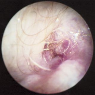
The external acoustic meatus and the skin on the pinna contain numerous adnexal structures and have a significant subcutaneous layer, which can respond to disease. Frequently, the stenotic portion of the ear is limited only to the external acoustic meatus (Figure 7-3). In that situation, the otoscope tip may be passed through the stenosis, revealing a normal vertical canal beyond it.
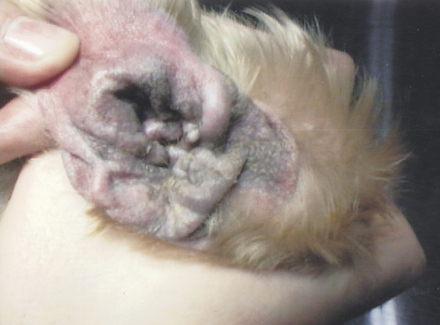
Figure 7-3 Hyperplastic epithelium on the concave pinna closes off the opening to the vertical ear canal.
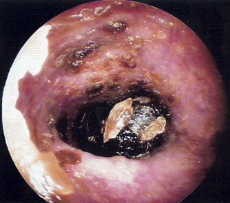
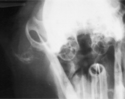
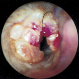
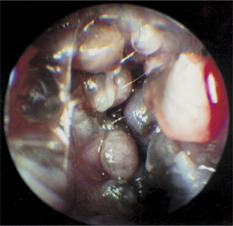
Inflammation and edema increase the thickness of the subcutaneous layer of the ear canal, leading to stenosis. Chronic otitis externa leads to progressive pathologic changes of the lining epithelium such as hyperkeratosis and hyperplasia (Figure 7-4). The marked thickening of the epithelial layer may significantly reduce ear canal diameter. Increases in the number and size of sebaceous glands (Figure 7-5) and dilated apocrine glands also reduce lumen diameter (Figure 7-6). In addition, pathologic changes that lead to calcification (Figure 7-7) and thickening of the auricular cartilage (especially in American Cocker Spaniels) or to fibrosis and formation of excessive granulation tissue resulting from chronic infection also lead to narrowing of the ear canal lumen (Figure 7-8). Tumors such as ceruminous adenocarcinoma may also occlude the ear canal lumen (Figure 7-9).
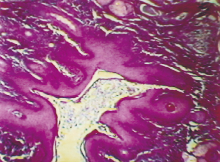
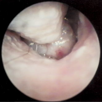
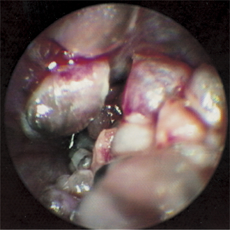
Figure 7-6 Ceruminous gland hyperplasia causes a stenosis in the vertical ear canal of this Cocker Spaniel.
Stenosis
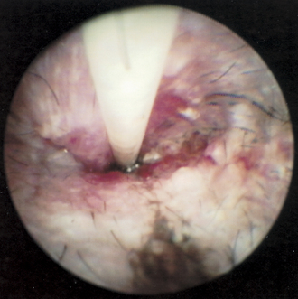
Stenosis presents a special problem in examination of the ear canal. Instruments cannot be inserted through the narrowed canal, and the integrity of the eardrum cannot be determined otoscopically. Occasionally, a short course of potent corticosteroids, applied topically, injected directly into the stenotic tissue, or administered parenterally, decreases the inflammatory infiltrates and reduces edema (Figure 7-10). In addition, corticosteroids decrease the amounts of glandular secretions, making them less viscous so that they are easier to flush out of the ear canal; they also reduce the secretion and dilation of ceruminous glands. If corticosteroids successfully increase the lumen diameter, adequate visualization of the ear canal and tympanic membrane becomes possible.
When medical therapy for stenosis is ineffective because of severe pathologic changes to the ear canal, surgical ablation of the vertical canal, horizontal canal, or both is required. Surgical treatment of stenotic ears relieves the pain associated with chronic otitis and allows the remaining ear canal to be ventilated.
Hair in Ear Canals
Routine plucking of hairs by groomers may not be necessary in a dog whose ears are normal, and it can sometimes be detrimental. Plucking the hairs with curved hemostats can create an inflammatory reaction that can predispose the ear to infection. In a dog that has recurrent ear infections and excessive hair growth in the ear canal, however, the hair should be routinely removed to prevent a mass of tangled hairs from blocking the ear canal lumen. Plucking the excessive hairs from the ears in patients predisposed to otitis externa is recommended for the prevention and management of otitis externa.
Stay updated, free articles. Join our Telegram channel

Full access? Get Clinical Tree


