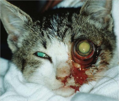
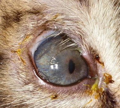
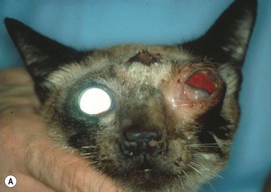
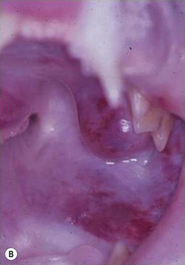
Chapter 57 Eyelids are composed of several different tissue types. The outer layer of haired skin covers a tarsal plate and musculature, which control the movement and positions of the eyelids. The inner layer adjacent to the globe is the palpebral conjunctiva, a thin, vascular tissue resembling a mucous membrane. The eyelids function to protect and support the globe. This is achieved by three methods: (1) they act as an initial defense against external dangers due to the presence of an active blink reflex; (2) they remove dirt and debris from the surface of the eye; and (3) they contribute portions of the tear fluid and distribute it across the ocular surface.1–5 The skin of the eyelids is thinner and more pliable than in other parts of the integument to allow for ease of movement. The thin subcutaneous tissue attaches the skin to the deeper orbicularis oculi muscle, which controls eyelid closure. Additional subcutaneous muscles allow for elevation of the upper eyelid (corrugators supercilii medialis medially and frontoauricularis laterally and the Müller muscle) and elongation of the palpebral fissure (retractor anguli oculi lateralis).1,3,4 Meibomian (or tarsal) glands are present along the margins of both the upper and lower eyelids and are responsible for producing the lipid fraction of the pre-ocular tear fluid. Their oily product exits through a series of openings that appear as little grey dots along the lid margin. Cilia, or eyelashes, are not normally present in the cat, although occasionally distichiasis, i.e., lashes that exit through the meibomian gland openings, are noted. Conjunctival goblet cells produce mucin, which is also a component of the tear fluid. The eyelids are innervated by branches of the facial nerve (sympathetic fibers and motor function) and by branches of the trigeminal nerve (sensory function). The majority of the vascular supply to the eyelids originates at the lateral and medial canthi from branches of the superficial temporal, the external ethmoidal, and infraorbital arteries. Lymphatic drainage from the eyelids is primarily to the parotid and mandibular lymph nodes.1,4 An extensive vascular supply is present in the orbit, most of which consists of various branches of the maxillary artery and their respective venous vessels which pass through the rostral alar foramen, the most ventrally situated foramen. The internal ophthalmic artery enters the orbit through the optic foramen with the optic nerve. The third, fourth, branches of the fifth, and the sixth cranial nerves, along with some autonomic fibers, enter the orbit through the orbital foramen and provide both sensory and motor innervation. The zygomatic salivary gland is small and lies close to the maxillary nerve near the ventral floor of the orbit. The nictitating membrane, or third eyelid, inserts in the ventromedial orbit. Adipose tissue fills the remaining space of the orbit and acts as a cushion to support the globe.1–3 When evaluating a cat for eyelid or periorbital conditions the signalment, in particular the age and breed of the cat, is relevant. A full history should be obtained from the owner in relation to how long the problem has been present, vaccination history, and history of concurrent disease such as rhinitis, history of trauma, etc. A full investigation of eye problems should include an intraocular examination and evaluation of vision.3,5 If a neoplastic condition is determined to be present, staging with thoracic and abdominal imaging and lymph node aspirates (see Chapter 14) may be indicated, depending upon the expected biologic behavior of the tumor. If surgical intervention in the orbit is anticipated, additional diagnostic procedures are recommended. B-scan ultrasonography of the orbit may provide useful information about the impact of the orbital disease on the globe and will often provide guidance for sampling of a lesion, but computed tomography (CT) and magnetic resonance imaging (MRI) are better options to delineate the extent and borders of the disease.6,7 A study in a cat that compared orbital echography, skull radiography, CT, and MRI to assess a mass involving the orbit showed that MRI was the only technique that delineated the entire border of the tumor.8 Prior to surgical intervention, histopathologic evaluation of a biopsy specimen is advised to facilitate surgical and postoperative treatment planning. Depending upon the exact location of the lesion within the orbit, sampling may be performed posterior to the bony orbital rim or via the oral cavity. Since the ventral orbital floor is bordered only partially by bone, the soft tissue posterior to the last molar may be incised in order to access the ventral orbit. A scalpel blade is used to incise the mucosa and a hemostat is gently and bluntly introduced through the incision, through the pterygoid musculature and the periorbita into the orbit for blind sampling. Expansive lesions may extend into the oral cavity and may be grossly appreciable upon examination of the mouth. Care must be taken to avoid damaging the maxillary artery as it courses along the orbital floor.1,2 If a diagnosis of a malignant process in the orbit is determined, the patient should have thoracic and abdominal imaging and potentially aspirates of the regional lymph nodes performed for staging purposes (see Chapter 14). Most eyelid disorders in cats will be the result of either congenital and developmental abnormalities or neoplastic diseases. Inflammatory conditions occur uncommonly and may be associated with infectious agents (Demodex, dermatophytoses, myiasis, FHV-1), autoimmune disorders (pemphigus, lupus, eosinophilic complex disease), or trauma and exposure to irritating compounds (drug reactions).3 Ankyloblepharon is fusion of the eyelids to one another. It is physiologic in the kitten until postnatal days 10–14.4 If the eyelids remain closed beyond this time, this indicates either incomplete development of the eyelids and periocular structures or more likely viral conjunctivitis (infection acquired from the queen either during parturition or during the first few days following birth) that has resulted in conjunctival adhesions or symblepharon. When an infection is introduced to the conjunctival space beneath the fused eyelids, neonatal ophthalmia results, which requires manual (premature) opening of the palpebral fissure. Often these kittens will have gross swelling of their orbits and eyelids and may have purulent discharge exiting through a small rent in the palpebral fissure. If the lids are left apposed when such an infection is present, the ocular surface will be irreparably damaged and vision will be significantly impaired if not lost completely. The incomplete formation of a portion of the eyelid is known as eyelid agenesis or coloboma (Fig. 57-1). It is usually associated with an absent eyelid margin.3 The severity of this condition varies dramatically among affected individuals.9 The majority of cases have absent lid tissue and margin along the temporal portion of the dorsal eyelid. Severe cases may have most of the upper lid missing, including the lateral canthus and portions of the lower eyelid as well. Mild cases may be managed medically with regular lubrication if minimal ocular irritation is present. Most cases, however, benefit from surgical reconstruction of the abnormal lids in order to provide greater coverage for the globe and to minimize discomfort from exposure and scarring that can impair vision. In many cases, ocular irritation will occur not only due to absent tissue and an impaired blink mechanism, but also as the result of trichiasis, in which hair from the face is deviated toward the cornea. Cats with eyelid agenesis often will have other ocular abnormalities including microphthalmia, cataract, retinal dysplasia, and keratoconjunctivitis sicca.9 A thorough ophthalmic examination should be performed to determine the extent of this congenital disorder. Figure 57-1 Eyelid agenesis in a nine-month-old domestic short-haired cat. The dorsotemporal eyelid margin is absent and trichasis is present. There is also keratitis, evidenced by corneal vascularization. The surgical approach to this condition will vary depending upon the severity. In some cases, simple apposition of the normal margins will suffice; other cases will require sliding advancement grafts (see Boxes 57-4 and 57-5) or grafts of tissue from either the lower eyelid or the commissure of the lip. Various surgical techniques have been employed.1,10–14 Most cases of eyelid malposition in cats are because of entropion, i.e., the inversion of the eyelid margin rolling toward the globe.3,15,16 Medial canthal entropion is almost universal in brachycephalic cats and is one of the causes of tear overflow and staining at the medial corners.1,3 In these cases, if irritation is minimal and the face can be kept sufficiently clean, surgical repair is not always necessary. In cats that develop full lid or lateral entropion, surgical correction is indicated.1,3,4,15 Most cats with entropion are only affected along the lower eyelid and occasionally at the lateral canthus (Fig. 57-2). Careful evaluation of the eyelid anatomy in affected cats is warranted since many cases have not only entropion, but also excessive length to their lower eyelid. In these cases shortening of the lower eyelid with a wedge resection may augment the repair.16 Ectropion, or eversion of the eyelid, is exceedingly rare in cats, and typically only results from cicatrix formation. Since eyelid neoplasms in cats are usually malignant, it is fortunate that they are relatively uncommon, occurring with significantly less frequency than cutaneous neoplasms elsewhere on the body.17,18 The prevalence of eyelid neoplasia increases with age in cats, but has not been found to correlate with breed or gender. The most common eyelid neoplasm in cats is squamous cell carcinoma. Other reported tumors include mastocytomas (Fig. 57-3), hemangiosarcomas and hemangiomas, fibrosarcomas, various neoplasms of glandular origin (adenomas, adenocarcinomas), basal cell carcinomas (Fig. 57-4), peripheral nerve sheath tumors, lymphomas, and others.3,17,18 Squamous cell carcinomas usually appear as ulcerated lesions along the eyelid margin with a crusting surface (Fig. 57-5). White cats seem to have a predilection for the development of this tumor.19 There is evidence for the role of actinic damage in the pathogenesis of this tumor. Squamous cell carcinomas are amenable to various forms of treatment and wide surgical excision is often curative, although grafting procedures are usually necessary to fill the resultant defect of excision. Most other types of feline eyelid neoplasm are also amenable to surgical excision, with variable recurrence rates.1,3,17 Mast cell tumors of the eyelids in cats generally respond favorably to surgical excision. A recent retrospective study described results in 33 cats with periocular mast cell tumors, and reported that recurrences were rare and affected cats had a median survival time of 945 days.20 Plesiotherapy with strontium-90 is another treatment option, either as a sole modality or in combination with surgical excision, that has excellent success rates.20,21 However, extensive lesions or disseminated disease may be best approached with systemic chemotherapy rather than excision or local radiation in cats.20 Cystic structures at the medial canthus that are benign and resemble apocrine hidrocystomas are occasionally noted in brachycephalic cats.18,22 Recurrence is common following local excision of these lesions, according to the authors’ experience and that of other veterinary ophthalmologists.18 The most common disorders of the feline orbit are neoplastic proliferations. They tend to be malignant (90%) and usually of epithelial origin.3 Squamous cell carcinomas are the most frequently reported orbital tumors in cats, followed by lymphoma (Fig. 57-6), fibrosarcoma and various bone tumors (Fig. 57-7).23 At least fifteen different tumor types have been reported within the feline orbit.23,24 Treatment strategies for each vary upon the extent of the lesion and the known or supposed biologic behavior of that type of tumor. Conservative surgical approaches to orbital neoplasms, which attempt to maintain the globe and vision, are usually associated with unacceptably high rates of tumor recurrence. Hence, the treatment of choice for orbital neoplasia is usually exenteration (see Box 57-7), which involves removal of the entire contents of the orbit including the globe. Orbitotomy techniques that spare the globe and seek to remove focal abnormalities within the orbit are rarely indicated in the cat and should be considered only in the case of small, well-defined lesions (preferably non-neoplastic or at least non-malignant). Advanced imaging of the head in cases of orbital neoplasia is strongly recommended because the incidence of concurrent orbital, nasal and sinus disease is high due to the close proximity of these spaces in cats. Figure 57-6 (A) A 12-year-old Siamese-cross with orbital lymphoma. (B) A bulging mass lesion and hemorrhage was noted posterior to the last upper maxillary molar upon oral examination of the cat. Unfortunately, because of the rigid nature of the orbital support, when a traumatic insult is sustained and the globe suffers injury, the great amount of force necessary to affect the globe usually results in significant damage, which may be vision or globe threatening. Proptosis, or the capture of the eyelid margins behind the equator of the globe (Fig. 57-8), usually with anterior displacement of the globe, is often associated with significant facial and orbital trauma and fractures of the skull and mandible.25 In most cases of traumatic proptosis in the cat, the affected globes are rendered non-visual and are so severely damaged that they require enucleation.1 The contralateral eye may also be blinded by damage to the optic chiasm from the stretching forces placed upon the affected globe.3 These cats are usually seriously injured and collapsed and should be triaged and treated first for shock, head-trauma, and concussive brain injury. If the globe can be salvaged, it should be kept well lubricated until the cat is stable enough to undergo anesthesia and a temporary tarsorrhaphy performed to provide for its protection.
Eyelids and orbit
Surgical anatomy
Eyelids
Orbit
General considerations
Eyelid evaluation
Diagnostic imaging
Histopathology
Surgical diseases of the feline eyelids
Ankyloblepharon
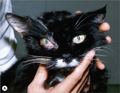
Eyelid agenesis
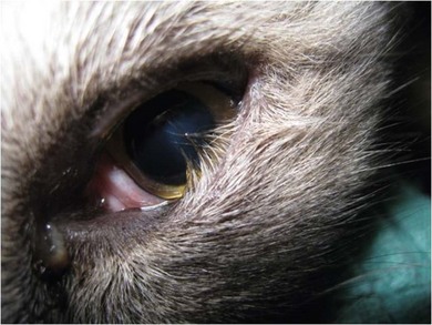
Entropion
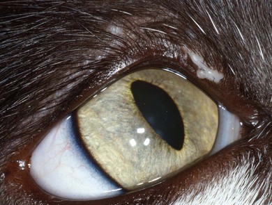
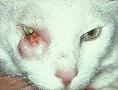
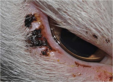
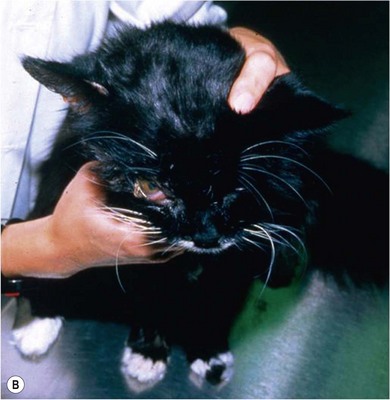
Eyelid neoplasia
Surgical diseases of the feline orbit
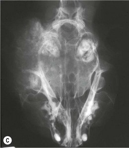
Orbital neoplasia
Proptosis
![]()
Stay updated, free articles. Join our Telegram channel

Full access? Get Clinical Tree


Eyelids and orbit