External Skeletal Fixation

Chapter 87External Skeletal Fixation


History and Development


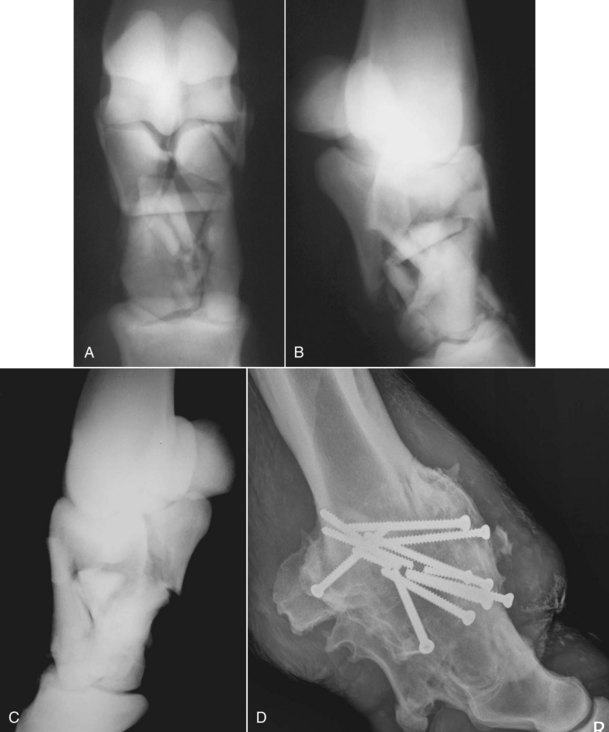
Using external skeletal fixation to treat fractures in the horse has only recently received some enthusiasm from equine surgeons. Although external skeletal fixation of fractures works well in small animals and has undergone periods of enthusiastic use, these same devices do not withstand the loads of weight bearing in an adult horse and therefore are not versatile enough to meet the needs of equine surgeons. Using transfixation pins in plaster, transfixation pins in fiberglass casts, or an external skeletal fixation device, has allowed salvage of some horses with difficult fractures that could not have been saved using internal fixation (Figure 87-1).


The incorporation of a walking bar cast, with transfixation pins placed in the bone above the fracture, was described in 1991 to manage fractures in horses and ponies.1 An overall success rate of 57% was reported for a variety of fractures in 35 horses and 21 ponies. The authors suggested that using transfixation pins incorporated into the cast material may help prevent fracture through these pin sites. Major complications of the technique included infection in nine horses or ponies, fracture through the bone or pin sites in six horses or ponies, and loss of circulation to the distal phalanx in two horses or ponies. Complications in the remaining seven nonsurvivors were not reported.1 Using in vitro tests, McClure and colleagues2,3 suggested that a walking bar was not necessary when using fiberglass casts and that divergent transfixation pins may be helpful in preventing fracture through the pin tract sites.


Although transfixation pins have been used successfully in fiberglass casts, this technique represents a compromise compared with classical external skeletal fixation. In horses managed with transfixation pin casts, wounds, skin, and pin sites must be covered in the cast, and at the time of every cast change, fractures are remobilized, allowing fracture collapse or shortening of the limb segment. In addition, pin loosening, pin breakage, and local infection at pin sites are common sequelae of this form of transfixation pinning, often necessitating pin replacement through new pinholes. The use of tapered-sleeve pins (TSPs) in a fiberglass cast has been reported.4 This technique increases the strength and durability of the pin and appears superior to use of unprotected pins alone, and fewer pins may be needed in the pin cast.4 This technique is used as an intermediate step after removal of the newly designed TSP external skeletal fixation device (ESFD) (see later).


The development of an equine ESFD in our laboratory had its origins before the first horse was treated in 1981, and we described the design and development of that device in the first 15 horses in 1986.5 The ESFD was designed to allow immediate full weight bearing in an adult horse and was used in horses with severely comminuted fractures of the distal aspect of the limb, in which internal fixation was not indicated or was impossible to carry out. A follow-up study including five additional horses along with further development of the device was published in 1992.6


Although the previous models of the ESFD proved the feasibility of using external skeletal pin transfixation in a frame device for immediate full weight bearing in an adult horse, there were substantial problems associated with the treatment itself. The original device used three unprotected 9.6-mm–diameter centrally threaded stainless steel pins loaded in bending within the intact bone (the third metacarpal bone [McIII]) above the fracture site, with two sidebars that connected the pins to a ground support through a base plate that was nailed to the horse’s hoof, much like a shoe. This allowed the forces of weight bearing to bypass a comminuted fracture with support through the pins, sidebars, and base. Six (22%) of 27 horses in which the original device was used developed fractures of the McIII through one of the pin holes (usually the proximal pin hole while wearing the device) either when wearing the device or shortly after removal of the device. Problems associated with catastrophic failure of the McIII through a pin hole after device removal were controlled by removing the device while the horse was standing rather than subjecting horses to the rigors of recovery from general anesthesia. After this modification there were no further incidents of McIII fracture after device removal (used in 12 horses without incident).


In 2001 we described a radical change of design of the transfixation pins that was aimed at reducing the incidence of these McIII failures through the pin holes.7 Several horses treated with this new design as well as horses treated with the old design were among a larger group of 64 horses with comminuted proximal phalangeal fractures reported in 2004.8


Jun 4, 2016 | Posted by in EQUINE MEDICINE | Comments Off on External Skeletal Fixation

Full access? Get Clinical Tree

Get Clinical Tree app for offline access