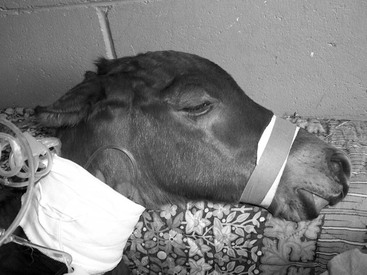
Kevin T. Corley, Jonna M. Jokisalo When a compromised neonatal foal is first presented, it is important to establish whether immediate emergency treatment is required. Three treatments that may need to be instituted in advance of obtaining a full history and clinical examination are fluid therapy, oxygen therapy, and cardiopulmonary resuscitation. The emergent need for one of these interventions is determined on the basis of quick assessment of the overall appearance of the foal, the heart rate and rhythm, the breathing rate and pattern, and mucous membrane color. Foals requiring immediate oxygen are likely to have low or high respiratory rates, may have respiratory distress, and may have cyanotic mucous membranes. Placing a normal neonatal foal in lateral recumbency reduces the arterial oxygen tension by approximately 10 mm Hg (1.3 kPa), which can make a substantial difference in a compromised foal. Therefore oxygen supplementation should be considered in foals in which restraint in lateral recumbency is needed, as during intravenous catheterization. Assessing whether a neonatal foal needs emergency fluids is not always straightforward. The clinical signs of hypovolemia (high heart rate, cold extremities, tachypnea, reduced jugular fill, poor pulse pressure) are not invariably present in hypovolemic foals. Foals that have not nursed for 3 hours or more are likely to be dehydrated, and foals that have not nursed for more than 6 hours are almost certain to be hypovolemic and require emergency fluids. The decision of whether to give immediate fluids is based on clinical signs, history, and a high index of suspicion. There are extremely few conditions in which giving 2 L of balanced electrolyte solution to a neonatal foal will cause harm, and in many cases it can significantly improve the foal’s chances of survival. Two (uncommon) conditions in which only small volumes of fluids should be given are uncontrolled hemorrhage and anuric renal failure. Key questions to ask in the history of a neonatal foal are: What signs is the foal showing? For how long has the foal had these signs? When was the foal born? Were any problems seen at the time of delivery? How was the foal’s behavior immediately after birth? How long was gestation? Has the mare had previous foals, and did they have problems? What is the parentage of the foal? For sick foals in the first couple of days of life, one of essentially two histories is likely. Either the foal looked well immediately after birth and then began to grow weak, or it was weak from birth. Both of these histories are common in foals with perinatal asphyxia syndrome and sepsis. Perhaps surprisingly, foals with conditions related to presumed birth trauma, such as ruptured bladder and diaphragmatic hernia, often appear normal after birth and only start showing clinical signs at 3 to 5 days. Likewise, foals with neonatal isoerythrolysis usually also are apparently initially normal and begin to show clinical signs at 1 to 7 days, most often 3 to 5 days after birth. The events at birth can profoundly affect the foal, with clinical signs usually becoming apparent within the first week of life but sometimes later. Prolonged birth (stage 2 lasting more than 40 minutes), premature placental separation, dystocia, and delivery by cesarean section are all risk factors for development of perinatal asphyxia syndrome, although the condition can also occur in foals in which no predisposing factors were recognized. Foals that require more than 60 minutes to stand or that do not nurse within the first 120 minutes after birth have significantly higher mortality and morbidity rates, compared with foals that stand and nurse inside these timepoints. The length of gestation is an important factor in the history and may direct early diagnostic testing to enable the owner to make an informed decision regarding whether to pursue treatment. However, gestation length alone is not sufficient grounds for determining prognosis because foals can be dysmature when born at term and or can be born early but have adequately mature organs and bone ossification to warrant giving a reasonable prognosis. The normal gestation length of mares ranges from 315 to 365 days, with a mean of 341 days. One critical factor in determining the prognosis for a foal that is born early is the state of the placenta and placental fluid. Perhaps counterintuitively, foals that are born from a “dirty” uterine environment are more likely to survive than those in which the placenta and uterine fluid appeared normal. This is because of precocious in utero maturation, in which increased corticosteroid concentrations in the mare induce hastened maturation of critical body systems in the fetus. The reproductive history of the mare can help in creation of a list of the likely differential diagnoses. If the mare has had previous foals, the length of prior gestations can help to determine whether the current pregnancy progressed normally. A small percentage of mares seems to repeatedly have foals with problems, although the problem may not be the same in every foal. Neonatal isoerythrolysis requires priming of the mare’s immune system by mismatching red blood cells, and is therefore rare (but not unheard of) in primiparous mares. Knowledge of a foal’s parentage provides an immediate indication of the foal’s potential future value and therefore the likely budget and expectations for treatment. The breeding can also give information as to the expected size of the foal (e.g., some stallions sire smaller foals than others) and any likely inherited flaws or characteristics. Clinical examination is typically divided into three phases. The first, the major body system assessment, was discussed previously. The second phase consists of a general overview of the foal and is often done subconsciously by experienced veterinarians from the moment they first see the foal. The third phase is a detailed head-to-toe examination of the foal. Attention to detail here is vital. There is little benefit in heroically saving a foal with severe sepsis if it will have permanently compromised vision because of undetected entropion. Much that determines both disease management and prognosis can be learned from the veterinarian’s general overview of the foal. The most important of these is the ability of the foal to stand and nurse unassisted. A foal that is not nursing will need both hydration and nutritional support. A foal that is unable to stand will require considerable nursing resources. It is also important to observe issues that will affect the future value of the foal; for the owner, these may be instrumental in deciding whether to invest in treatment. In many instances, the owner may not have seen the foal and is relying on the veterinarian to find any defects before embarking on treatment. Even if the owners are present, they may be so concerned about the primary disease that they miss other issues that will become a problem in the future. Issues that can be observed during the general overview include limb conformation, scoliosis or other vertebral column defects, and parrot mouth. More obvious defects, such as hydrocephalus or congenital skin lesions, are likely to be part of the presenting complaint. Although in general it is best to conduct a head-to-tail examination, the cardiovascular system is of such priority that it is usually assessed first. A brief examination of this system (heart rate and mucous membrane color) is included in the initial major body system assessment. At that point, it is usually decided what other assessments of the cardiovascular system are necessary. At a minimum, in addition to being checked for heart rate and mucous membrane color, the foal should be examined for jugular vein filling, cold extremities, skin tenting, and pulse pressure. Pulse pressure is simply the difference between systolic and diastolic arterial pressure and does not give information about the mean pressure (e.g., if the diastolic pressure is low, the pulse pressure can feel normal despite a low mean arterial pressure). In a recumbent foal presented to a hospital, indirect mean arterial pressure should also be measured. Many similarities exist in the head-to-tail physical examination between the foal and adult horse. However, there are also many differences, and some parts of the animal that would normally only receive a cursory examination in an adult horse unless there is a specific indication warrant detailed evaluation in the foal. One example of this is the aural pinnae. Start with the mouth and oral mucous membranes. The mucous membranes should be pink and moist, with capillary refill time less than 2 seconds. The mucous membranes can indicate changes in hydration, alterations in microcirculation, and the coagulation system. Dry or tacky mucous membranes are associated with inadequate hydration. Purple mucous membranes indicate poor oxygenation, which can be caused by a lung problem or heart defect that causes right-to-left shunting of blood, whereas red, injected mucous membranes can be seen with peripheral vasodilation. Yellow or icteric mucous membranes are most commonly a sign of neonatal isoerythrolysis. However, sepsis, neonatal hyperbilirubinemia, impaired hepatic function, equine herpesvirus type 1 infection, iron toxicosis, and meconium impaction can all increase circulating bilirubin concentrations and therefore also manifest as jaundice. Petechiae can also form in the oral mucous membranes and can be a result of sepsis or of the ulcerative dermatitis, thrombocytopenia, and neutropenia syndrome. The tongue should be examined for signs of infection, such as candidiasis. Cleft palate can usually only be detected on physical examination if the foal is comatose or anesthetized, or if it involves the hard palate (a rare occurrence). A cleft soft palate is usually detected endoscopically. Brachygnathia (parrot mouth), wry nose, congenital oral tumors such as hemangiosarcoma, and other abnormalities can also be detected at this time. The nares should be checked for airflow during exhalation because unilateral choanal atresia may not be clinically obvious in neonatal foals. Confirmation of choanal atresia is made by endoscopy. As in adult horses, the nares should also be checked for discharge. There are two primary differential diagnoses for milk coming out of the nares during nursing: milk regurgitation and physical interference with swallowing, such as that caused by a cleft palate or subepiglottic cyst. Milk regurgitation appears to be an organ-specific form of perinatal asphyxia syndrome in which there is a temporary swallowing defect. It is treated by preventing nursing while feeding the foal by nasogastric tube. In most cases, swallowing ability is restored in 48 to 96 hours. If left untreated or caught late, these foals will develop aspiration pneumonia. Milk regurgitation must be differentiated by endoscopy from physical problems affecting swallowing. Milk or saliva escaping from the nose and mouth can occur both with esophageal obstruction and with esophageal ulceration. Serous nasal discharge can develop with neonatal viral diseases, such as equine influenza (especially in an unvaccinated population), and mucopurulent discharge is often a sign of bacterial disease. The pupillary light reflex is universally present in newborn foals but can appear slightly sluggish, compared with the reflex in adult horses. The menace response only develops in the first week of life. In one study of 26 foals, 1 foal had a menace response on day 1 of life, most foals had a menace response by day 7, and all foals had a menace response by the ninth day. Entropion is relatively common in neonatal foals, especially compromised foals, and should be corrected if observed. Sepsis can manifest in the eyes as hypopyon (pus in the anterior chamber) or hyphema (blood in the anterior chamber of the eye). The eyes should also be checked for size because congenital microophthalmia can occur. The conjunctival mucous membranes can develop color changes similar to those in the mouth, but the changes are sometimes more obvious in the conjunctiva. The sclera is often the first place where subtle jaundice is appreciated. A domed head and floppy ears are signs of prematurity in the foal (Figure 172-1); premature foals can also have fine, silky hair coats. The inside of the ears (aural pinnae) are the classically described place to detect petechiation and ecchymosis in the foal (Figure 172-2). However, petechiae can be fairly subtle in this location, and other mucous membranes should also be examined for these lesions.
Evaluation of the Compromised Neonatal Foal
Major Body System Assessment (Triage)
History
Clinical Examination
General Overview
Cardiovascular Assessment
Examination From Head to Tail
Head and Neck
< div class='tao-gold-member'>
![]()
Stay updated, free articles. Join our Telegram channel

Full access? Get Clinical Tree


Evaluation of the Compromised Neonatal Foal
Chapter 172
Only gold members can continue reading. Log In or Register to continue
