CHAPTER 13 Equipment
Radiographs are images of the shadows made when x-rays (electromagnetic waves of pure energy that generate both electrical and magnetic fields) incompletely and differentially penetrate imaged objects. X-rays that are able to pass through the object interact with the film or sensor, while those that are absorbed or deflected by radiodense objects do not. This creates a shadow image on the film or sensor that consists of a “gray scale” of black through white that corresponds to x-rays traveling through the object uninterrupted (black) through complete blocking (white) and shades of gray between. This scale can have many different levels of gray (long scale) with very subtle differences between them, or it can have very few gradations of gray (short scale) that are much more distinct.
Dental Radiograph Machines
X-ray equipment for general radiography in most veterinary facilities consists of a stationary tube head suspended over a patient-positioning table. Dental radiographs can be made using this equipment using extraoral technique (see Chapter 12). However, a dedicated dental x-ray machine greatly facilitates creating dental and oral radiographs. Intraoral technique with a dedicated dental x-ray machine provides high-quality images without leaving the dental operatory. The purchase cost is a modest investment with a fast return when used as it should be.
GENERATOR
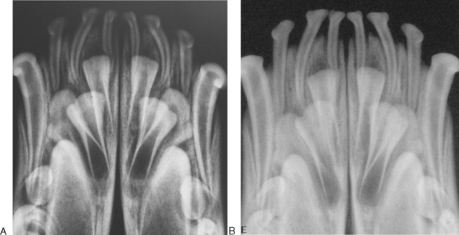
The generator sends a precise quantity and quality of energy to the tube head, stimulating the firing of electrons toward a target (anode) in the tube head. The quantity of electrons is determined by the amperage and exposure time (milliampere-seconds, or mAs), and their acceleration energy toward the target is determined by the applied voltage (kVp). The target takes the delivered quantity of electrons and converts their energy into x-ray photons. Some dental x-ray units may have set kVp and milliamperage (mA) values, while others allow the operator to select an mA setting between 7 and 15 mA and a kVp setting between 60 and 90 kVp. Higher kVp and lower mAs make fewer x-rays with higher penetrating ability. In contrast to this, using a low kVp and a high mAs tends to produce images using more x-rays but with less penetrating ability. A balance needs to be struck between images that are pleasing to view with high contrast and very black-and-white character, and images that are more challenging to interpret due to the additional shadows and more shades of gray but also contain more diagnostic information (Figure 13-1). All units allow the operator to select an exposure time, either directly or indirectly. The exposure time may be in fractions of a second, or it may be in “pulses” that are 1/60th of a second.
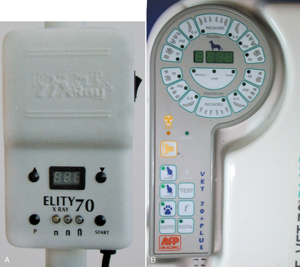
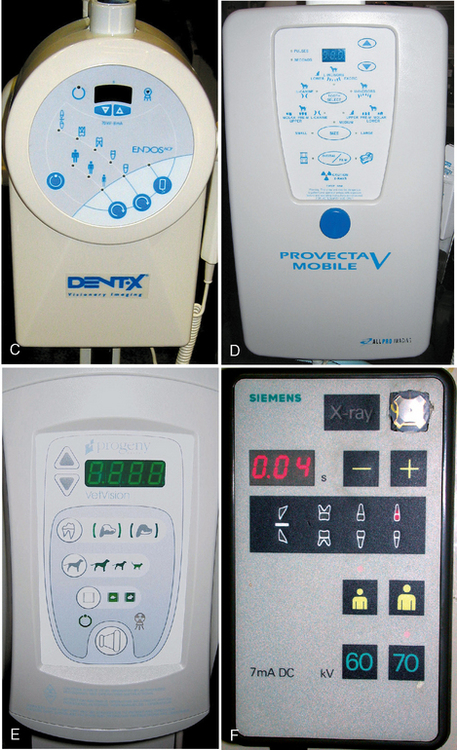
CONTROL UNIT AND INTERFACE
The user interfaces vary between units. The simplest units with a set kVp and mA have only a power button, a digital timer exposure control, and an exposure button (Figure 13-2, A). Others do not allow direct selection of exposure time, instead using an anatomical interface that requires the operator to select the size and species of the patient and the specific tooth being radiographed (Figure 13-2, B). Many now allow either anatomical selection or direct timer selection (Figure 13-2, C–F). Those with the anatomic interface also have some way of setting the equipment to the operator’s specific site variables (cone length, film speed, or sensor type) that affect the amount of radiation needed for diagnostic images. The exposure button on the control unit needs to be pressed throughout the exposure. Removing pressure mid-exposure interrupts the generation of x-rays and usually generates a machine error code to warn the operator of the underexposure. This is designed as a safety precaution.