Carrie J. Finno
Equine Neuroaxonal Dystrophy
Equine neuroaxonal dystrophy (NAD) is a common neurologic disorder that affects many breeds of horses. The disease is characterized by symmetrical ataxia, abnormal base-wide stance at rest, and proprioceptive deficits in all limbs, signs that usually develop by 6 to 12 months of age. Affected foals often have low serum vitamin E concentrations. Neuroaxonal dystrophy is clinically indistinguishable from equine degenerative myeloencephalopathy (EDM); the only difference between these disorders is the location of axonal degeneration within the central nervous system. These conditions appear to have a genetic basis, with clinical expression in genetically predisposed foals being influenced by dietary vitamin E. The genetic defect underlying development of NAD and EDM is currently unknown.
On the basis of data from postmortem studies, NAD/EDM was rated the second most common cause (24% of cases) of spinal cord disease in horses by Mayhew and colleagues at Cornell University in 1978 and ranked second in the causes of spinal ataxia at the University of Montreal from 1985 to 1988. At present, a clinical diagnosis is supported by normal cervical radiographs or myelography, negative results of equine protozoal myeloencephalitis (EPM) testing, and low serum vitamin E concentrations, specifically α-tocopherol. Careful histologic evaluation of the central nervous system at postmortem examination is currently the only way to definitively diagnose NAD or EDM. Neuroaxonal dystrophy/equine degenerative myeloencephalopathy easily goes unrecognized because an antemortem diagnostic test is not available, and postmortem histopathologic changes can be subtle.
Nomenclature
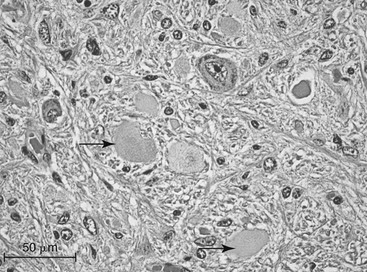
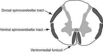
Equine NAD and its closely related counterpart EDM are clinically indistinguishable and also resemble other causes of symmetrical spinal cord ataxia, such as cervical vertebral compressive myelopathy (CVCM). The distinction between NAD and EDM depends mainly on the distribution of the histologic lesions. Histologic lesions in both NAD and EDM consist of neuronal and axonal degeneration, and clinical manifestations of the two diseases are also distinguished chiefly by the distribution of the lesions. With equine NAD, lesions are primarily confined to specific nuclei of the brainstem, namely the nucleus cuneatus lateralis (Figure 89-1), nucleus cuneatus medialis, and nucleus gracilis, and additional specific nuclei within the thoracic segments of the spinal cord (nucleus thoracicus). Histologic lesions of EDM extend into specific regions of the white matter of the spinal cord, namely the dorsal and ventral spinocerebellar tracts and ventromedial funiculi of the cervicothoracic part of the spinal cord (Figure 89-2). In Quarter Horses with histopathologic changes consistent with EDM, lesions consistent with NAD have also been detected. Therefore the term NAD is used to encompass the primary disease process, whereas EDM is considered a more severe variant of NAD.
Etiology
Role of α-Tocopherol
The following evidence suggests that vitamin E, specifically α-tocopherol, plays an important role in the development of NAD and EDM in genetically predisposed foals. Compared with foals sired by an unaffected stallion, those from an EDM-affected stallion have significantly lower plasma α-tocopherol concentrations, and α-tocopherol supplementation of these foals during the first year of life or of both pregnant dams and foals lowers both the overall incidence and the severity of NAD and EDM. Although vitamin E clearly plays a role in these susceptible families, low serum α-tocopherol is not consistently reported in all cases of NAD or EDM. Furthermore, when serum α-tocopherol concentrations are low, it is unrelated to abnormal vitamin E absorption, suggesting that there is a difference in vitamin E requirements or metabolism in affected or susceptible animals. Overall, there is strong evidence that NAD and EDM are inherited disorders, and research to date suggests that the serum concentration of α-tocopherol acts as an environmental modifier to determine the overall severity of the NAD or EDM phenotype.
Inheritance
Strong evidence supports a genetic basis for NAD and EDM. Foals from dams that have had an EDM-affected foal are 25 times as likely to develop EDM as foals from other dams. In a prospective breeding trial, 9 of 15 offspring from affected Morgan horses were affected, compared with 0 of 22 offspring from control horses. Clustering of cases among related horses provides further strong evidence of a genetic basis for NAD and EDM. Based on the breeding study, a dominant mode of inheritance with variable expression or a polygenic mode of inheritance was suggested; however, there have been no outcrosses to verify this theory.
In a family of clinically phenotyped Quarter Horses with NAD/EDM sharing the same underlying risk factor of a deficiency in α-tocopherol, heritability was recently estimated at 70%. Complex segregation analysis excluded both an X-linked pattern of inheritance and a fully penetrant (i.e., 100% of cases with the genetic mutation display the disease phenotype) autosomal dominant mode of inheritance. Therefore it appears that NAD and EDM may be inherited as an incompletely penetrant autosomal dominant trait, with vitamin E, specifically α-tocopherol, concentrations during the first year of life modifying the overall phenotype of affected horses.
Comparatively, the clinical and histologic findings in horses with NAD or EDM resemble ataxia with vitamin E deficiency (AVED) in humans. Human AVED is caused by various mutations in the α-tocopherol transfer protein gene (TTPA), which encodes for the protein responsible for vitamin E transport in the liver and incorporation of α-tocopherol, a main component of vitamin E, into very low density liposomes for transport throughout the body. The author’s investigations have excluded TTPA as a candidate gene for NAD in a family of Quarter Horses. Because the only strong candidate gene for NAD/EDM has been excluded, a genome-wide association study and further gene expression analyses are currently underway to provide insight into the genetic cause of NAD and EDM in Quarter Horses and other breeds.
Stay updated, free articles. Join our Telegram channel

Full access? Get Clinical Tree


