CHAPTER 10 Endocrine, Nutritional, and Miscellaneous Hair Coat Disorders
Endocrine disorders
Pituitary pars intermedia dysfunction
Pituitary pars intermedia dysfunction (PPID, Cushing disease, Cushing syndrome, hirsutism) is the most common endocrinopathy of the horse—one of the most common diseases of horses and ponies 15-years old and older—and has been estimated to affect 15-30% of aged horses.2–5 Clinical signs are caused by increased blood concentrations of the hormones secreted by an adenoma or hypertrophied cells in the pars intermedia of the pituitary gland (hypophysis).
It is hypothesized that PPID is primarily a dopaminergic neurodegenerative disease.2,3,5,19 Dopamine-containing neurons are particularly sensitive to oxidative stress, and markers of oxidative stress are increased in the hypothalamus of horses with PPID. There is no evidence of the systemic accumulation of oxidative stress marker or of deficiencies in antioxidant capacity in horses with PPID.20 Horses with PPID have increased expression of interleukin-8, perhaps indicating that inflammatory dysregulation may contribute to the development of PPID.24 It is currently believed that PPID results from loss of normal dopaminergic inhibitory control, eventuating in the classic endocrine processes of hypertrophy, hyperplasia, and, rarely, adenoma formation. For reasons that are currently unknown, there is a decrease or loss of the neurotransmitter dopamine in the innervation of the pars intermedia. The concentrations of dopamine and dopamine metabolites are up to ninefold less in the pars intermedia of horses with PPID as compared with age-matched control horses.5,19,20 There is decrease in pituitary dopaminergic nerve terminals and a loss of periventricular dopaminergic neurons in horses with PPID.5,19,20 The function of melanotrophs in the pars intermedia is normally inhibited when dopamine is released from nerve endings extending from the hypothalamus. Loss of inhibition is followed by increased synthesis and secretion of various peptides. Over time there is hypertrophy and hyperplasia of the pars intermedia melanotrophs. In most cases of PPID, the pars intermedia is enlarged because of hypertrophy and hyperplasia, with progression to true adenoma being a late and uncommon event.
The normal equine pituitary gland produces a variety of closely related peptides by cleavage of a common precursor protein known as proopiomelanocortin (POMC).2,5 The pars distalis processes POMC to adrenocorticotropin (ACTH), β-lipotropin (βLPH), and α-lipotropin (αLPH), whereas in the pars intermedia, the main POMC-derived peptides are α-melanocyte stimulating hormone (α-MSH), β-melanocyte-stimulating hormone (β-MSH), corticotropin-like intermediate lobe peptide (CL1P), and β-endorphin (β-END). In normal horses, the pars intermedia produces only about 2% of the ACTH. Horses with PPID have elevated concentrations of these POMC-derived peptides in pituitary tissue and blood and a loss of the diurnal (circadian) rhythm of cortisol secretion. ACTH release from the pars distalis is under neuroendocrine control from the hypothalamus, mediated via corticotropin-releasing hormone (CRH) and arginine vasopressin and is subject to negative feedback by cortisol. In contrast, pars intermedia POMC-peptide secretion is under dopaminergic control and does not appear to be influenced by plasma cortisol. Loss of dopamine results in increased expression and plasma concentrations (up to 50-fold) of POMC-derived peptides (including α-MSH, β-END, CLIP, and ACTH).3-5,24
The alteration in POMC-derived hormones contributes to adrenocortical dysfunction by at least two mechanisms.2–5 Release of ACTH in horses with PPID is much greater than that observed in clinically normal horses and, although it is not as pronounced as the release of other POMC-derived peptides, it is sufficient to stimulate adrenocortical steroidogenesis. Also, other POMC-derived peptides can potentiate the actions of ACTH. Thus, a small increase in ACTH concentration, coupled with a large increase in potentiating peptides (e.g., MSH and β-END), can contribute to melanotroph-mediated adrenocortical dysfunction. Increased cortisol secretion results in insulin resistance that, in turn, is associated with dyslipidemia and laminitis.
Other causes of equine hyperadrenocorticism are rare. Two cases, one caused by a pituitary meningioma and the other by an adrenocortical adenoma are reported.4 Classical hyperadrenocorticism due to exogenous steroids (iatrogenic disease) has not been reported.
The pathogenesis of the clinical signs seen in PPID are complex and, in many instances, undocumented.2–5 Weight loss and muscle weakness and atrophy are presumably due to the catabolic effects of glucocorticoids. Type 2 myofiber atrophy present in muscle biopsy specimens from horses with PPID is consistent with glucocorticoid-associated atrophy.5 The occurrence of fat pads is thought to be due to fat redistribution under the influence of glucocorticoids. Increased susceptibility to bacterial and fungal infections and poor wound healing are due to the immunosuppressive and antiinflammatory effects of glucocorticoids. Laminitis is hypothesized to be associated with the enhancement of the vasoconstrictor potency of endogenous biogenic amines by glucocorticoids. Glucocorticoid excess is also believed to be the cause of frequent low blood concentrations of thyroid hormones, abnormal estrus cycles, and rarely, fractures.
Behavioral changes (e.g., lethargy, docility, decreased response to painful stimuli) could be due to high levels of β-END in blood and cerebrospinal fluid or pressure of the pituitary lesion on the hypothalamus.2,4,5 The pressure of an enlarging pituitary mass can also cause central blindness, thermoregulatory disturbances, and seizures.
The most puzzling clinical abnormality is the hypertrichosis, which has been postulated to be due to: (1) increased adrenocortical production of androgens, (2) increased secretion of α-MSH, or (3) pressure of the pituitary lesion on thermoregulatory areas of the hypothalamus.2,4,5 Although the hair coat abnormality has classically been referred to as hirsutism, this is inappropriate. Hirsutism refers to hair growth in women in areas of the body where hair growth is under androgen control and in which normally only postpubescent males have terminal hair growth (e.g., mustache, beard, chest).1 The hair coat abnormality in PPID is properly called hypertrichosis, which specifically refers to hair density or length beyond the accepted limits of normal for a particular age, race, or sex.1 Hyperhidrosis may be caused by pressure of the pituitary lesion on thermoregulatory areas of the hypothalamus or the dramatic hair coat change.2–5
Clinical features
Horses that present with many or most of the classical historical and physical features of PPID make for a straightforward clinical diagnosis.2–5 However, the clinical signs often develop sequentially and in random order over a variable period of time (months to years). Signs may wax and wane. The presenting complaint for horses with PPID is frequently not directly related to the pituitary dysfunction, and evidence to suggest pituitary dysfunction is often revealed by a carefully developed history and thorough physical examination.
PPID is generally a disease of elderly horses (average 20-years old, range 7- to 42-years old). There are no clear breed predilections, but ponies and Morgans may be at increased risk.2–5 There is probably no sex predilection.
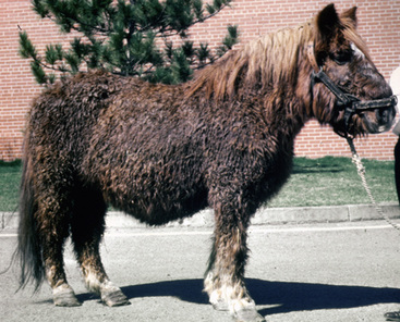
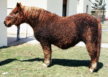
Hypertrichosis (“hirsutism”) is the classic and most consistent clinical sign associated with PPID. With the exception of the spontaneous hypertrichosis seen in the Bashkir curly breed, there is no other condition in which horses develop a long, curly, hair coat.2–5 Hence, acquired hypertrichosis in an aged horse or pony can be considered a positive diagnostic test result for PPID.3,5,8 The hair coat is typically long, thick, shaggy, and curly, often most noticeably on the limbs (Figs. 10-1 and 10-2). The mane and tail are typically unaffected. Early signs may be delayed shedding in spring, often followed by early regrowth of winter coat in fall. Some horses exhibit incomplete shedding, with long hairs persisting in the jugular groove, under the shins, the belly, and the legs, or in a few round patchy areas over the entire body. Before generalized hypertrichosis is appreciated, affected horses often develop longer hairs on the legs, ventrum, and under the chin. Some horses with early PPID develop transient alopecia with shedding, commonly confined to the head. In exceptional cases, there may be a coat color change. The skin may be normal, dry and scaly, or greasy.
Horses with PPID are increasingly susceptible to skin infections (especially dermatophilosis, staphylococcal folliculitis, abscesses, and draining tracts). Hyperhidrosis—usually episodic but occasionally persistent—is seen in up to 60% of the cases. This can lead to matting of the hair coat into thick tufts. Rarely, papular and nodular xanthomas are present over the thorax.4
Weight loss, muscular weakness, and muscular atrophy are present in up to 88% of the cases. The epaxial, lumbar, and abdominal muscles are particularly affected, often leading to a pot-bellied and sway-backed appearance.2–5 The pendulous abdomen may be confused with weight gain. Muscle wasting can also result in an increased prominence of the croup, tuber coxae, and tuber sacrale regions. Fat pads may develop on the body, especially in the supraorbital, tailhead, lumbar, neck, and scapular regions. Bilateral nonpainful periorbital swelling in the absence of ocular disease is very suggestive of PPID.3
Polydipsia and polyuria are seen in 39-76% of the cases and are usually associated with diabetes mellitus and/or diabetes insipidus.2–5 Changes in behavior and attitude (lethargy, docility, dullness, drowsiness, somnolence, decreased responsiveness to painful stimuli) are common (15-82% of cases).2–5
Laminitis is seen in 24-82% of affected horses and may be present in the absence of other classic clinical signs.2,3,5,13 Often all four feet are affected, and horses suffer repeated bouts of laminitis that is refractory to conventional therapy. Intractable pain is probably the most common reason for euthanasia. Any horse older than 7-years old that develops laminitis in the absence of a clear inciting cause should be tested for PPID.3
Other signs that may be seen in horses with PPID include polyphagia or anorexia, buccal ulcers, episodic tachypnea and tachycardia, bacterial or mycotic infections (conjunctivitis; sinusitis; pneumonia; joint or tendon sheath infections; abscesses in teeth, feet, pharynx, mandible, and lung), increased susceptibility to parasitism, neurologic abnormalities (blindness, ataxia, seizures), infertility, abnormal estrus cycles, delayed wound healing, rare skeletal problems (fractures, hypertrophic osteopathy), colic, and rare pancreatitis.2-6,19a
Diagnosis
When many of the classical abnormalities are present, a presumptive diagnosis of PPID is justified based on history and physical examination. Because acquired hypertrichosis in an aged horse or pony is believed to be pathognomonic for PPID, its presence can be considered a positive diagnosis.2,3,5,8 One must not confuse the hypertrichosis of PPID with normal long winter hair coats. Retention of long winter hair coat can be seen in horses with chronic illnesses and nutritional deficiencies.4 However, in early cases, wherein only one or two of the clinical signs (e.g., weight loss, behavioral change, neurologic disturbance, laminitis, infection, infertility) are present, the differential diagnosis can be quite lengthy.2–5
Results of routine laboratory testing are variable, often reflective of disease chronicity, and never used to confirm or refute the diagnosis.2–5 Typical, fully developed cases frequently have insulin-resistant hyperglycemia; glucosuria; varying combinations of relative or absolute neutrophilia, lymphopenia, and eosinopenia (stress leukogram); elevated plasma insulin concentrations; and low serum thyroxine (T4) and triiodothyronine (T3) concentrations. Mild anemia is often present. Urine specific gravity may be normal or low. Horses with chronic infections may have leukocytosis and nonregenerative anemia. Occasional horses have elevated serum liver enzyme concentrations, and others have lipemia, hypercholesterolemia, and hypertriglyceridemia.
Adrenal function tests
As is often the case when multiple diagnostic protocols are described for a given disease, no single method has emerged as being consistently superior to any other for the diagnosis of PPID. Diagnostic testing is complicated by the fact that there is marked seasonal variation in many test results.* Concentrations of endogenous hormones such as ACTH and α-MSH increase in the fall in the northern hemisphere. In addition, results of the dexamethasone suppression tests are less reliable in the fall. Thus, diagnostic testing is best done before June or after October in the northern hemisphere. The effect of season on diagnostic testing needs to be more extensively studied using large numbers of horses and ponies in diverse geographic locations.
In the past, the postmortem histologic evaluation of the pituitary gland was often used as the gold standard for the evaluation of antemortem diagnostic tests.2,3,21 However, it has been reported that there is no agreement among pathologists on the histopathologic definition of PPID.21 Hence, the use of histologic evaluation as a gold standard is inappropriate.
Blood cortisol
Baseline blood cortisol concentrations in normal horses approximate 1-13 mcg/dL.2,5 Baseline blood cortisol concentrations are not affected by breed, age, gender, or pregnancy. However, important considerations to keep in mind when interpreting blood cortisol levels include the following: (1) different laboratories may differ in their normal and abnormal values; (2) stress (exercise, hypoglycemia, anesthesia, surgery, severe disease, transport, hospitalization, blood taking, unaccustomed to environment or routine) can markedly elevate blood cortisol levels; (3) episodic daily secretion of cortisol occurs; and (4) single measurements of blood cortisol are of no value in the diagnosis of PPID.12,12a Because of the endogenous rhythmic and episodic fluctuations, the exogenously provoked fluctuations in baseline cortisol concentrations, and the overlap with baseline concentrations found in healthy horses, blood cortisol measurements are highly unreliable for the diagnosis of PPID. Basal cortisol concentrations in horses with PPID may be low, normal, or high.
ACTH stimulation test
Two commonly used ACTH stimulation test procedures for the horse are as follows: (1) plasma or serum cortisol determinations are made before and 8 h after the intramuscular (IM) injection of 1 International Unit/kg of ACTH gel, or (2) plasma or serum cortisol determinations are made before and 2 h after the intravenous (IV) injection of 100 International Units of synthetic aqueous ACTH. By either procedure, normal horses will double to triple their basal cortisol levels, whereas horses with PPID will have exaggerated responses. The sensitivity of the ACTH stimulation test in the diagnosis of PPID varies from 70% to 79%.2,4,5 Thus, it does not consistently discriminate between normal and PPID horses. If the ACTH stimulation test results are combined with the measurement of plasma ACTH concentrations, sensitivity was reported to be 100%.4
Dexamethasone suppression test
Older literature indicated that the dexamethasone suppression test was not a sensitive indicator of adrenocortical function in horses.4 In horses, dexamethasone does not have the suppressive effect seen in dogs and humans, presumably because the hypersecretion of ACTH is from the pars intermedia (rather than the pars distalis) and is relatively insensitive to glucocorticoid negative feedback. These observations were based on dexamethasone suppression tests performed by measuring plasma or serum cortisol before and 6 h after the IM administration of 20 mg dexamethasone.
The overnight dexamethasone suppression test was reported to be 100% sensitive and 100% specific and was considered the gold standard test for the diagnosis of PPID.2–5 Basal blood cortisol concentrations are measured prior and 19-24 h following the administration of 40 mg (0.04 mg)/kg dexamethasone IM or IV. Dexamethasone suppression tests have occasionally been avoided or discouraged in horses with PPID because of the hypothetical risk of causing laminitis. However, no side effects have been reported with the overnight protocol. Starting a dexamethasone suppression test at 9 AM or 9 PM gave the same results. After an overnight dexamethasone suppression test, normal horses will have postdexamethasone cortisol concentrations of less than 1 mcg/dL, while horses with PPID will have concentrations of over 1 mcg/dL. More recently, it has been reported that overnight dexamethasone test results are less reliable in the fall in the northern hemisphere,3,8,12,32 and that the same horses can have test results positive for PPID at one moment in time and normal at another.3,18,27 When overnight dexamethasone suppression tests were performed monthly in aged horses without clinical signs of PPID, all horses had normal test results in November through April, but often had abnormal (positive) test results in June through October.5a,32
Plasma ACTH
Basal plasma ACTH concentrations in normal horses approximate 5-37 pg/mL.2–5 Blood for ACTH assays is typically drawn in ethylenediaminetetraacetic acid (EDTA) tubes, and the plasma is separated within 3 h and frozen at −20 °C in a plastic tube. Once frozen, the plasma can be kept for up to 1 month before being assayed. Samples of frozen plasma should be shipped to the appropriate laboratory on dry ice overnight to ensure that they do not thaw during shipment.
Plasma ACTH concentrations are reported to be sensitive and specific for the diagnosis of PPID in horses and ponies, but are not elevated in all patients.3,4,29 Plasma ACTH concentrations over 27 pg/mL (ponies) and over 50 pg/mL (horses) are “strongly supportive” of the diagnosis of PPID. It has been reported that the simultaneous performance of an ACTH stimulation test and basal plasma ACTH concentrations was 100% accurate for the diagnosis of equine PPID.4 More recently it has been reported that plasma ACTH concentration in healthy horses and ponies are markedly increased (positive for PPID) in the fall.2,3,5a,12
TRH stimulation test
Thyrotropin-releasing hormone (TRH) stimulates pars intermedia melanotrophs, resulting in increased concentrations of plasma cortisol, α-MSH, and ACTH.2,3,17 An increase in blood cortisol of 90% or greater above baseline concentration 15-30 min after the IV administration of 1 mg of TRH is consistent with a diagnosis of PPID. The sensitivity and specificity of the TRH stimulation test are inferior to the overnight dexamethasone suppression test.2,3,14,25 TRH can be difficult to procure and is expensive. TRH is generally well-tolerated, with occasional bouts of trembling, yawning, lip-smacking, flehmen response, salivation, urination, defecation, miosis, tachycardia, and tachypnea reported.3,5,14
Other tests
The combined dexamethasone suppression/ACTH stimulation test often yields ambiguous results.4
The combined dexamethasone suppression/TRH stimulation test was reported to have a sensitivity, specificity, positive predictive value, and negative predictive value of 88%, 76%, 71%, and 90% respectively.14 Plasma cortisol concentrations are measured pre- and 24 h post-IV injection of 0.04 mg/kg dexamethasone and 30 min post-IV injection by 1 mg TRH (which is given 3 h after the dexamethasone injection). This protocol is expensive, not superior to more widely studied diagnostic protocols, and would presumably be affected by season and geographic location.
Although the mean urinary corticoid:creatinine ratio is significantly higher in PPID than in normal horses, there is much overlap in test results, resulting in both false-negative and false-positive test results.2–5
Basal plasma insulin concentrations, the glucose tolerance test, and the insulin response test have good sensitivity, but are only applicable to horses with hyperglycemia.2-5,22 Although insulin-resistant hyperglycemia is commonly seen in PPID horses, stress-induced hyperglycemia in normal horses may result in glucose concentrations of the same order of magnitude.2-5,8 In addition, hyperglycemia with normal basal insulin concentrations has been reported.4 Horses with equine metabolic syndrome may exhibit insulin resistance.5,6 Furthermore, ponies often have a relative insensitivity to insulin compared with horses.4
Horses with PPID have a diurnal pattern of serum insulin concentrations, with highest values present at noon (12 PM).22 In a study of horses with PPID treated with trilostane,22 lower serum insulin concentrations (less than 62 μu/mL) at the beginning of therapy were associated with improved survival as compared to higher concentrations (over 188 μu/mL). In that study, serum insulin concentrations in normal horses were reported to range from 5.4 to 36 μu/mL.
Plasma concentrations of α-MSH are presumably a more pars intermedia-specific measurement.3,17,18 As is the case for plasma ACTH concentrations, plasma α-MSH concentrations are reported to be markedly increased in normal horses and ponies in the fall in the northern hemisphere.18 TRH injections markedly increase plasma α-MSH concentrations in normal horses, horses with PPID, and pars intermedia explants.17 Samples for plasma α-MSH measurement must be kept frozen until assayed. No commercial test is currently available.
Plasma ACTH concentration responses to domperidone (3.3 mg/kg orally [PO] peripheral dopamine D2 receptor blocker) were correlated with pituitary histomorphometry in horses with PPID.28 Results suggested the protocol may be useful for the diagnosis of PPID.
In a very small number of horses, salivary concentrations of cortisol in PPID horses were higher than those in normal horses.4 Injections of ovine CRH were reported to increase plasma and salivary concentrations of cortisol in normal horses.30
Imaging
Several imaging protocols (computerized tomography, magnetic resonance imaging, ventrodorsal radiography with contrast venography) have been described for PPID, but none has received general acceptance.2-4,26 Such techniques require general anesthesia and the availability of specialized equipment. In addition, the rate of false-negative studies (masses less than 2 mm in diameter) and false-positive studies (nonfunctional pituitary lesion) is unknown. An age-related increased prevalence of lesions in the pars intermedia and pars distalis in clinically normal horses and ponies has been reported.33
Stay updated, free articles. Join our Telegram channel

Full access? Get Clinical Tree


