Verena K. Affolter
Draft Horse Lymphedema
Chronic progressive lymphedema (CPL) is a disabling disorder of draft horse breeds, characterized by progressive swelling of the distal portions of the limbs, hyperkeratosis, marked dermal fibrosis, and formation of skin folds and nodules. These lesions are typically complicated by recurrent bacterial and parasitic infections, which further impair drainage of lymph. Equine CPL resembles primary lymphedema in humans, also referred to as elephantiasis verrucosa nostra. Primary lymphedema in humans is an inherited disorder, which has been mapped to various genes, including FOXC2.
Equine CPL has been best characterized in Shires, Clydesdales, and Belgian draft horses. Lesions compatible with CPL also develop in Gypsy Vanners, English Cobs, and Friesians, but the condition has not been studied as extensively in these breeds. Although Percheron horses in the United States seem mostly unaffected, CPL has been observed in this breed in Europe. A chronic progressive and debilitating disorder, CPL leads to severe disfigurement, loss of use of the horse because of lameness, and premature euthanasia.
Clinical Signs
Clinical signs of CPL vary with the stage of the disease and the complicating presence of secondary infections. Both forelimbs and hindlimbs are affected, but the lesions tend to be more pronounced in the hindlimbs.
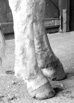
First lesions can be seen as early as 2 years of age. Typically, early mild signs remain unnoticed because the heavy feathering obscures the early pitting edema and mild hyperkeratosis. Removal of the feathering reveals the pitting edema and mild skin rippling on the skin surface (Figure 124-1). Lesions are most prominent in the fetlock and pastern area. At this stage, secondary infections may become established and initiate inflammation of the skin and subcutis. Any inflammatory event will further enhance the disturbed lymph flow. With progression of the pitting edema, a clear definition of the cannon bone, flexor tendons, and fetlock joint contours is lost, and the extremities appear more cone shaped.
Secondary infections mostly include infections with staphylococcal species and Chorioptes mites, but occasionally may include Dermatophilus congolensis and other bacteria. Chorioptes spp infections elicit marked pruritus, which is evidenced by stamping of the feet. Excoriations may develop secondary to the constant scratching of limbs.
Prolonged pitting edema initiates fibrosis and induration of the skin and subcutis, which further enhances the decrease in lymph flow. In addition to increased circumference of the distal legs, firm folds and nodules are first observed in the palmar and plantar area of the pastern region. Folds and nodules progressively enlarge over time (up to several centimeters in depth or diameter, respectively). Eventually, both folds and nodules develop proximal to the fetlock and may extend all the way up to the carpus and tarsus. The skin surface is severely hyperkeratotic, and excessive scaling is seen.
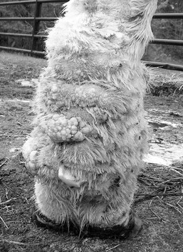
Owners often only become alarmed at a later stage of the process, when lesions become obvious despite heavy feathering. With increasing induration from the lymphedema, lymph drainage and appropriate tissue perfusion are remarkably impaired, resulting in erosions and ulceration of skin folds and nodules. The secondary bacterial and parasitic infections recur more frequently and are more severe. The skin surface oozes, bleeds, and forms crusts. The lesions may be not only pruritic but also painful, and many horses become very reluctant to have the limbs touched. The marked induration with prominent folds and nodules, in particular when inflamed, eventually interfere with normal gaits, and larger folds are subject to self-traumatization by the opposite limb (Figure 124-2). Persistent infections can progress into deeper tissues and induce lymphangitis and swelling of the entire limb. The deep skin folds with the oozing surface are ideal niches for maggot infestation.
Horses with CPL often have poor hoof quality, characterized by brittle, chipped walls with splits and cracks. The coronary band is markedly hyperkeratotic, and hooves are broad and deformed. Repeated bouts of thrush and deep hoof abscesses are commonly seen, and some horses develop laminitis.
Etiology
The exact etiology of equine CPL has not been identified. Similar to primary lymphedema in humans, equine CPL is associated with impaired circulation and lymph drainage, which results in impaired skin barrier function. The latter, together with the occlusive environment created by the heavy feathering, sets the stage for recurrent bacterial (Staphylococcus spp and Dermatophilus congolensis) and parasitic (Chorioptes spp) infections. Elastin is crucial for effective functioning of lymphatic vessels and appropriate lymph drainage. Affected Shires, Clydesdales, and Belgian horses have clear evidence of altered elastin metabolism and degradation of elastin, identified by altered morphologic features of the elastin network on histology and by increased levels of circulating antielastin antibodies. Within affected breeds, it is a challenge to find completely unaffected horses among animals older than 10 years of age. Moreover, certain familial lines of draft horses are more affected than others. This clearly indicates a genetic background to this disorder, which would reflect the situation in humans with primary lymphedema. However, the genetic predisposition of equine CPL has not been characterized to date; identified nucleotide polymorphism in the equine FOXC2 gene was not consistently present among affected horses tested in one report.


